. 2015 Apr;34(4):296-302.
doi: 10.1089/dna.2014.2627. Epub 2015 Feb 18.

Upregulation of Sestrin2 expression protects against macrophage apoptosis induced by oxidized low-density lipoprotein

Affiliations

Upregulation of Sestrin2 expression protects against macrophage apoptosis induced by oxidized low-density lipoprotein

Hong-Juan Hu et al. DNA Cell Biol. 2015 Apr.

Abstract

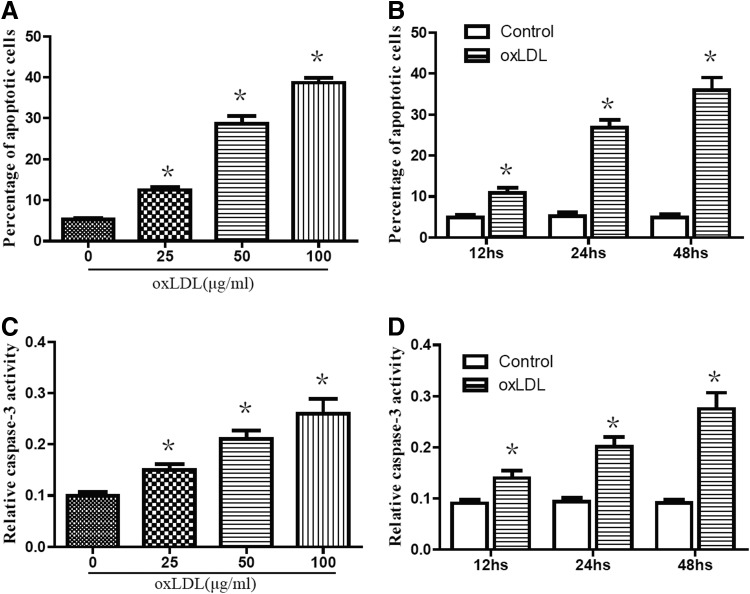
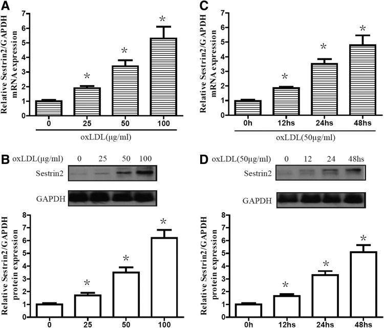
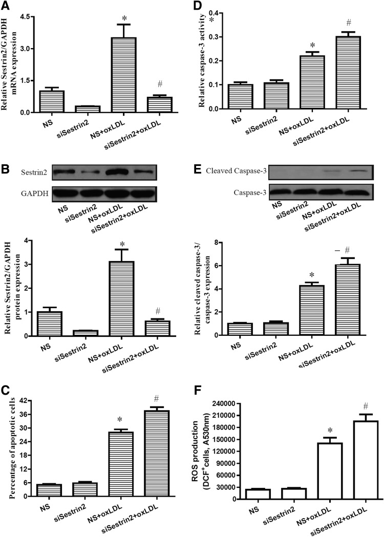
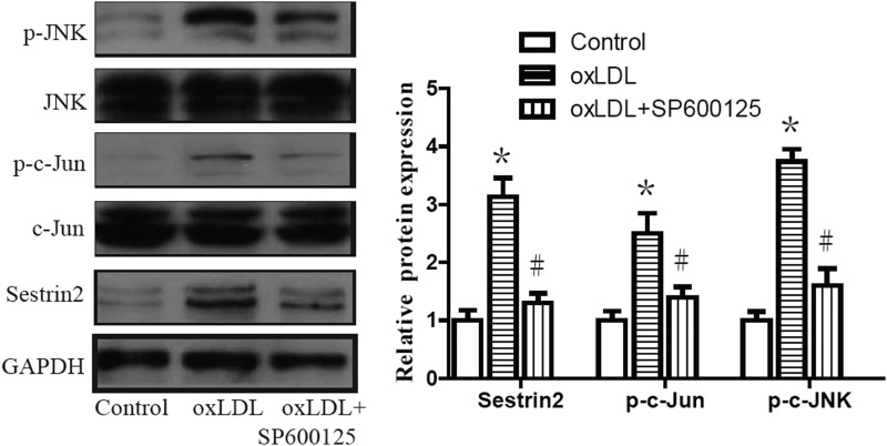
Sestrin2 is involved in a different cellular response to stress conditions. However, the function of Sestrin2 in the cardiovascular system remains unknown. In the present study, we tested whether Sestrin2 has a beneficial effect on macrophage cell apoptosis induced by oxidized low-density lipoprotein (oxLDL). We found that oxLDL induces expression of Sestrin2 in RAW264.7 cells in a time-dependent and dose-dependent manner. We also found that knockdown of Sestrin2 using small RNA interference promotes cell apoptosis and reactive oxygen species production induced by oxLDL. In addition, our results show that the c-Jun NH(2)-terminal kinase (JNK)/c-Jun pathway is activated by oxLDL. Inhibiting the activity of the JNK pathway abolishes the increase of Sestrin2 induced by oxLDL. These findings suggest that the inductive effect of Sestrin2 is mediated by the JNK/c-Jun pathway. Our results indicate that the induction of Sestrin2 acts as a compensatory response to oxLDL for survival, implying that stimulating expression of Sestrin2 might be an effective pharmacological target for the treatment of lipid-related cardiovascular diseases.


Figures

<b>FIG. 1.</b>
FIG. 1.
Oxidized low-density lipoprotein (oxLDL) treatment increased cell apoptosis in RAW264.7 cells. (A, C) RAW264.7 cells were stimulated with oxLDL at various concentrations for 24 h. (B, D) RAW264.7 cells were stimulated with oxLDL at indicated doses (50 μg/mL) for varying periods of time. Cell apoptosis was determined by FACS analysis with PE-Annexin-V staining. Caspase-3 activities were measured as described in the Materials and Methods section. Data are presented as the mean±SEM of at least three independent experiments. *p<0.05 versus nontreated control.
<b>FIG. 2.</b>
FIG. 2.
oxLDL induced the expression of Sestrin-2 in RAW264.7 cells. (A, B) RAW264.7 cells were stimulated with oxLDL at various concentrations for 24 h. (C, D) RAW264.7 cells were stimulated with oxLDL at indicated doses (50 μg/mL) for varying periods of time. Protein levels of Sestrin-2 were determined by western blot analysis. mRNA levels of Sestrin-2 were determined by real-time polymerase chain reaction (RT-PCR). Data are presented as the mean±SEM of at least three independent experiments. *p<0.05 versus nontreated control.
<b>FIG. 3.</b>
FIG. 3.
Sestrin2 downexpression induced apoptosis and reactive oxygen species (ROS) production of RAW264.7 cells in presence of oxLDL. RAW264.7 cells were transfected with NS or siSestrin2. At 48 h after the transfection, cells were treated with 50 μg/mL oxLDL for 24 h. (A, B) Expression of Sestrin-2 was determined by RT-PCR and western blot analysis. (C) FACS analysis with PE-Annexin-V staining revealed that Sestrin2 downexpression induced apoptosis. (D) Caspase-3 activities were measured as described in the Materials and Methods section. (E) Cleaved caspase-3 expression was detected by western blot analysis. (F) Intracellular ROS production was determined by the fluorescence probe 5-chloromethyl-2′,7′-dichlorodihydrofluorescein diacetate, acetyl ester (CM-H2DCFDA). Data are presented as the mean±SEM of at least three independent experiments. NS nonspecific RNA, siSestrin2 Sestrin2 small RNA interference. *p<0.05 versus NS control; #p<0.05 versus NS+oxLDL group.
<b>FIG. 4.</b>
FIG. 4.
c-Jun NH(2)-terminal kinase (JNK)/c-Jun mediates oxLDL-induced upregulation of Sestrin2. RAW264.7 cells were treated with oxLDL with or without the presence of 25 nM of JNK inhibitor SP600125 for 24 h. Western blot analysis revealed that the levels of p-JNK, p-c-Jun, and Sestrin2 are increased by oxLDL. Data are presented as the mean±SEM of at least three independent experiments. *p<0.05 versus nontreated control; #p<0.05 versus oxLDL-treated group.

References

    1. Bae S.H., Sung S.H., Oh S.Y., Lim J.M., Lee S.K., Park Y.N., Lee H.E., Kang D., and Rhee S.G. (2013). Sestrins activate Nrf2 by promoting p62-dependent autophagic degradation of Keap1 and prevent oxidative liver damage. Cell Metab 17,73–84 - PubMed
    1. Bonawitz N.D., Chatenay-Lapointe M., Pan Y., and Shadel G.S. (2007). Reduced TOR signaling extends chronological life span via increased respiration and upregulation of mitochondrial gene expression. Cell Metab 5,265–277 - PMC - PubMed
    1. Budanov A.V., and Karin M. (2008). p53 target genes sestrin1 and sestrin2 connect genotoxic stress and mTOR signaling. Cell 134,451–460 - PMC - PubMed
    1. Budanov A.V., Sablina A.A., Feinstein E., Koonin E.V., and Chumakov P.M. (2004). Regeneration of peroxiredoxins by p53-regulated sestrins, homologs of bacterial AhpD. Science 304,596–600 - PubMed
    1. Budanov A.V., Shoshani T., Faerman A., Zelin E., Kamer I., Kalinski H., Gorodin S., Fishman A., Chajut A., Einat P., Skaliter R., Gudkov A.V., Chumakov P.M., and Feinstein E. (2002). Identification of a novel stress-responsive gene Hi95 involved in regulation of cell viability. Oncogene 21,6017–6031 - PubMed

Publication types

MeSH terms

LinkOut - more resources