. 2015 Feb 18:6:6243.
doi: 10.1038/ncomms7243.

A cnidarian homologue of an insect gustatory receptor functions in developmental body patterning

Affiliations

A cnidarian homologue of an insect gustatory receptor functions in developmental body patterning

Michael Saina et al. Nat Commun. .

Abstract

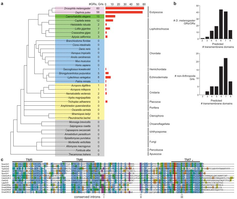
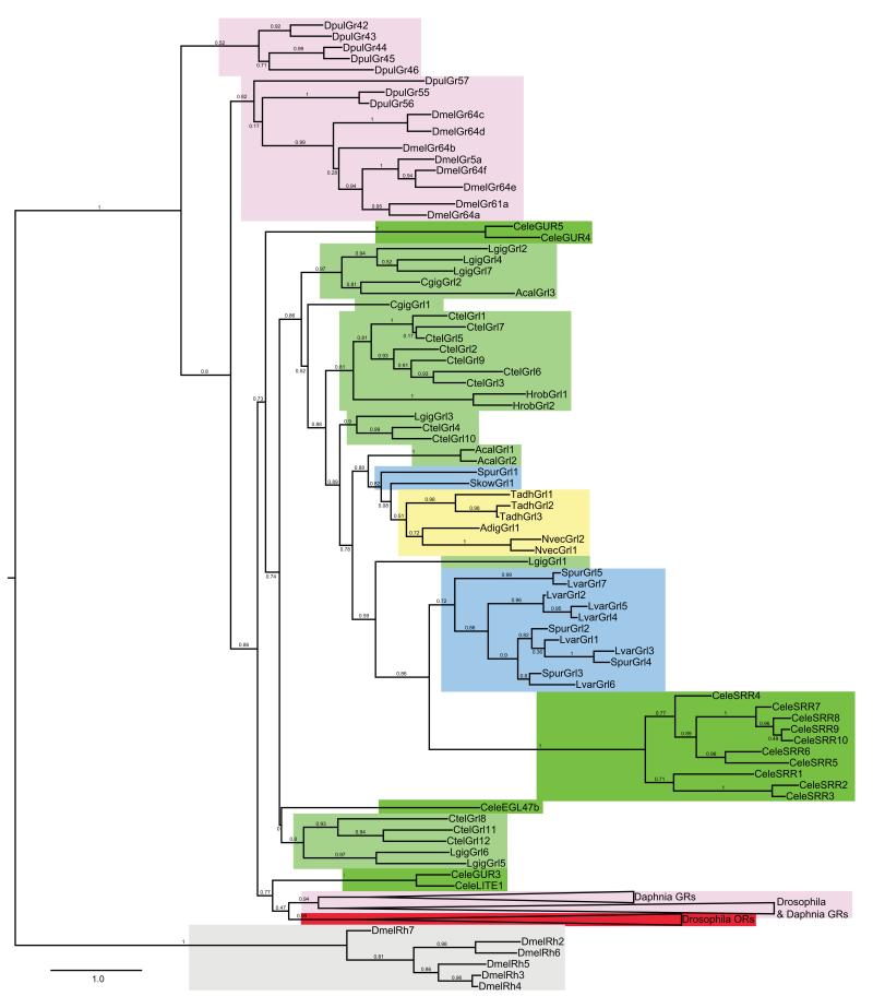
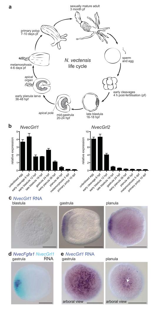
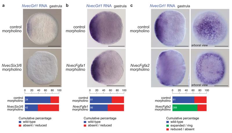
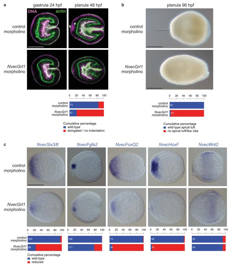
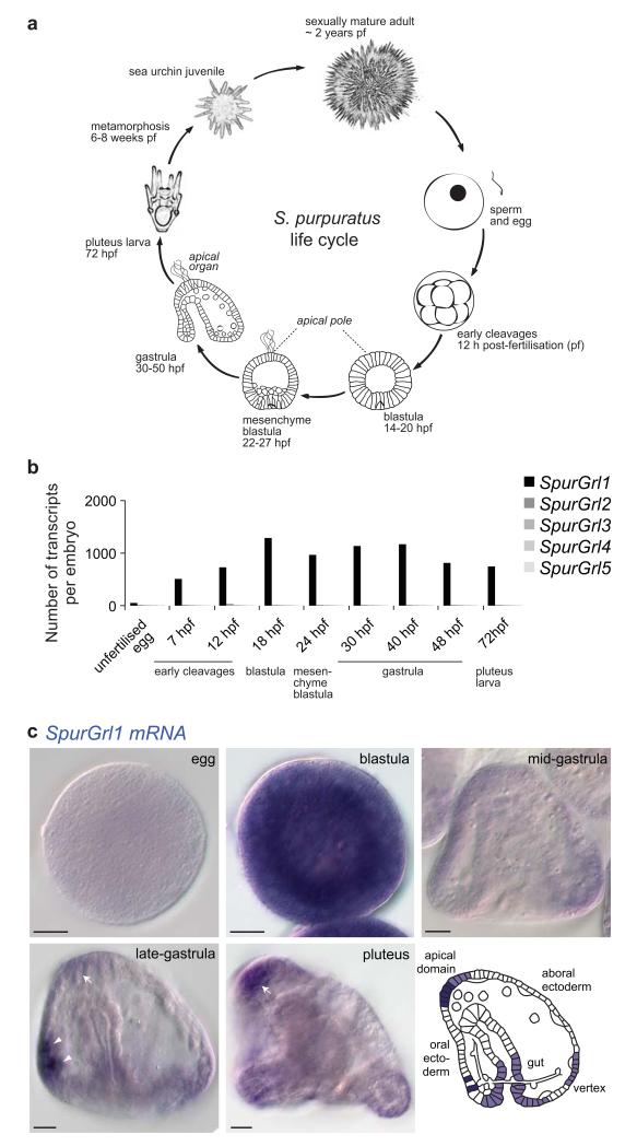
Insect gustatory and odorant receptors (GRs and ORs) form a superfamily of novel transmembrane proteins, which are expressed in chemosensory neurons that detect environmental stimuli. Here we identify homologues of GRs (Gustatory receptor-like (Grl) genes) in genomes across Protostomia, Deuterostomia and non-Bilateria. Surprisingly, two Grls in the cnidarian Nematostella vectensis, NvecGrl1 and NvecGrl2, are expressed early in development, in the blastula and gastrula, but not at later stages when a putative chemosensory organ forms. NvecGrl1 transcripts are detected around the aboral pole, considered the equivalent to the head-forming region of Bilateria. Morpholino-mediated knockdown of NvecGrl1 causes developmental patterning defects of this region, leading to animals lacking the apical sensory organ. A deuterostome Grl from the sea urchin Strongylocentrotus purpuratus displays similar patterns of developmental expression. These results reveal an early evolutionary origin of the insect chemosensory receptor family and raise the possibility that their ancestral role was in embryonic development.


Figures

Figure 1
Figure 1. Identification of Gr-like (Grl) genes
(a) Summary of the GR/Grl repertoires identified in the genomes of selected arthropods (pink), non-arthropod Ecdysozoa (dark green), Lophotrochozoa (light green), Deuterostomia (blue) and non-Bilateria (yellow). An unscaled tree showing the phylogenetic relationships between these species is illustrated on the left; relationships among the different non-bilaterian phyla is unresolved, with the exception of the Cnidaria, which are the closest sister group to Bilateria. Related genes were identified in multiple species of nematode worms but, for simplicity, only C. elegans is shown. (b) Computational predictions of the number of transmembrane domains found in D. melanogaster GRs and ORs (top) and Grls (bottom). Grl sequence fragments (lacking start/stop codons) were excluded from this analysis. (c) Alignment of the C-terminal regions of select insect GRs and ORs, C. elegans GURs and Grls. Predicted transmembrane (TM) domains are indicated with horizontal lines. The asterisk marks a conserved tyrosine residue important for ion conduction in insect ORs. The arrowheads below the alignment indicate the positions of three phase 0 ancestral GR introns (inferred from analysis of D. melanogaster GRs and ORs) that are conserved in Grls: intron I is conserved in phase and position in NvecGrl1, NvecGrl2, TadhGrl2, SpurGrl1, CeleGUR2, CeleGUR3, CtelGrl2, LgigGrl3; intron II is conserved in phase and position in NvecGrl1, NvecGrl2, TadhGrl2, SpurGrl1, CtelGrl2, and LgigGrl3. Intron III is conserved in all sequences.
Figure 2
Figure 2. Phylogenetic analysis of Grl, GR and OR genes
Maximum likelihood tree showing the relationships between select arthropod GRs/ORs, C. elegans GURs and all Grls (except the short sequence fragments predicted in P. miniata and A. millepora), colour-coded as in Fig. 1a. Drosophila Rhodopsins are included as an outgroup; although it remains unclear whether these two different families of heptahelical membrane proteins share a common ancestor, they clearly belong to a separate clade. For clarity, branches for large clades of D. melanogaster GRs/ORs or D. pulex GRs have been collapsed. The scale bar represents the number of amino acid substitutions per site.
Figure 3
Figure 3. N. vectensis Grls are expressed during early development
(a) Schematic of the life cycle of the sea anemone N. vectensis. Dark grey shading in blastula, gastrula and early planula stages indicates the endoderm. (b) Quantitative RT-PCR analysis of the temporal expression of NvecGrl1 and NvecGrl2 during nine developmental time-points. Data from three biological replicate samples (mean ± s.e.m.) are shown. (c) RNA in situ hybridisation using a riboprobe against NvecGrl1 on whole mount N. vectensis at three developmental stages. Lateral views, with the arboral side on the left, are shown for all specimens in this and subsequent figures, unless otherwise noted. (d) Two-colour RNA in situ hybridisation using riboprobes against NvecGrl1 (light blue) and NvecFgfa1 (dark blue) on whole mount N. vectensis embryos. (e) RNA in situ hybridisation using a riboprobe against NvecGrl1 on whole mount N. vectensis at two developmental stages showing an apical view, which reveal the formation of a ring-like distribution of transcripts at the planula stage. The apical organ forms from the unstained cells inside the ring (white arrow). Scale bars in (c-e) = 100 μm.
Figure 4
Figure 4. Regulation of N. vectensis Grl1 expression by the apical patterning network
(a) RNA in situ hybridisation using a riboprobe against NvecGrl1 on whole mount N. vectensis embryos injected with either control or NvecSix3/6 morpholino oligonucleotides. Quantification of the phenotypes is shown below. Note that NvecGrl1 transcripts are relatively weakly detected and a fraction of control morpholino injected animals did not exhibit staining. The n is shown in white within each bar in this and all equivalent graphs. (b) RNA in situ hybridisation using a riboprobe against NvecGrl1 on whole mount N. vectensis embryos injected with either control or NvecFgfa1 morpholino oligonucleotides. Quantification of the phenotypes is shown below. (c) RNA in situ hybridisation using a riboprobe against NvecGrl1 on whole mount N. vectensis embryos injected with either control or NvecFgfa2 morpholino oligonucleotides. In NvecFgf2 morphants, note the precocious formation and larger size of the ring of NvecGrl1 transcripts around the aboral pole (compare with Fig. 3e). Quantification of the phenotypes is shown below. Scale bars in (a c) = 100 μm.
Figure 5
Figure 5. N. vectensis Grl1 is required for arboral pole patterning
(a) Morphological phenotypes of N. vectensis embryos injected with either control (control#1) or NvecGrl1 (NvecGrl1#1) morpholino oligonucleotides, in which DNA (magenta) and the actin cytoskeleton (green) are labelled with TO-PRO-3 and Alexa Fluor 488 Phalloidin, respectively. The arrow marks the group of nuclei at the aboral pole that have translocated to a more basal position (towards the right of the image) in control but not NvecGrl1 morphants; this results in these cells forming a small indentation of the apical ectoderm (arrowhead). Scale bar = 100 μm (applies to all images). Quantification of the phenotypes is shown below. (b) Bright-field images of living 4 day planulae derived from N. vectensis embryos injected with either control (control#1) or NvecGrl1 (NvecGrl1#2) morpholino oligonucleotides revealing the failure in apical tuft develops in NvecGrl1 morphants. Quantification of the phenotypes is shown below. Scale bars = 100 μm. (c) RNA in situ hybridisation using probes against marker genes in animals injected with either control (control#1) or NvecGrl1 (NvecGrl1#1) morpholino oligonucleotides. Scale bar = 100 μm (applies to all images). Quantification of the phenotypes is shown below.
Figure 6
Figure 6. Early developmental expression of an S. purpuratus Grl
(a) Schematic of the life cycle of the sea urchin S. purpuratus. (b) Quantitative RT-PCR analysis of the temporal expression of the five SpurGrl genes during nine developmental time points. Data are represented as number of transcripts per embryo and represent the average of four technical replicates in each of two independent biological replicate samples (see Supplementary Table 3). (c) RNA in situ hybridisation using a riboprobe against SpurGrl1 on whole mount S. purpuratus of five developmental stages. All the embryos represent lateral views: as indicated in the schematic, the oral ectoderm is on the left and the apical domain on the top in gastrula and pluteus specimens. The arrows mark the expression of SpurGrl1 in the apical domain (where the apical organ will form), while the arrowheads mark a pair of presumptive neurosecretory cells in the oral ectoderm. Scale bars = 20 μm.

References

    1. Robertson HM, Warr CG, Carlson JR. Molecular evolution of the insect chemoreceptor gene superfamily in Drosophila melanogaster. Proc. Natl. Acad. Sci. USA. 2003;100(Suppl 2):14537–14542. - PMC - PubMed
    1. Hallem EA, Dahanukar A, Carlson JR. Insect odor and taste receptors. Annu. Rev. Entomol. 2006;51:113–135. - PubMed
    1. Benton R. Chemical sensing in Drosophila. Curr. Opin. Neurobiol. 2008;18:357–363. - PubMed
    1. Nei M, Niimura Y, Nozawa M. The evolution of animal chemosensory receptor gene repertoires: roles of chance and necessity. Nat. Rev. Genet. 2008;9:951–963. - PubMed
    1. Vosshall LB, Stocker RF. Molecular Architecture of Smell and Taste in Drosophila. Annu. Rev. Neurosci. 2007;30:505–533. - PubMed

Publication types

MeSH terms

LinkOut - more resources