. 2015 Feb 18:6:6244.
doi: 10.1038/ncomms7244.

Multiplex CRISPR/Cas9-based genome editing for correction of dystrophin mutations that cause Duchenne muscular dystrophy

Affiliations

Multiplex CRISPR/Cas9-based genome editing for correction of dystrophin mutations that cause Duchenne muscular dystrophy

David G Ousterout et al. Nat Commun. .

Abstract

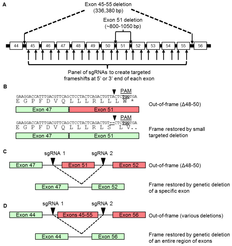
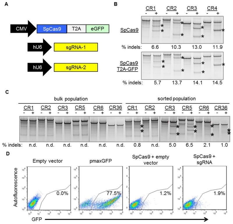
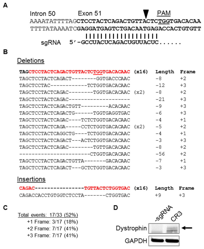
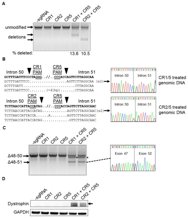
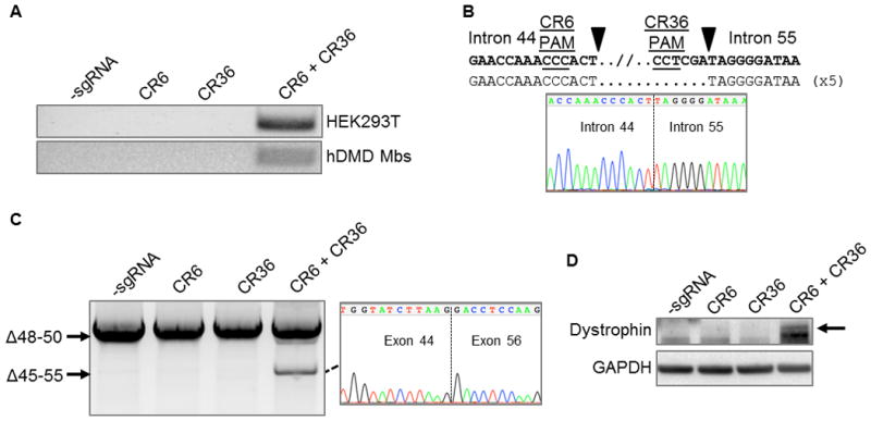
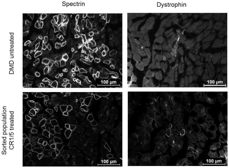
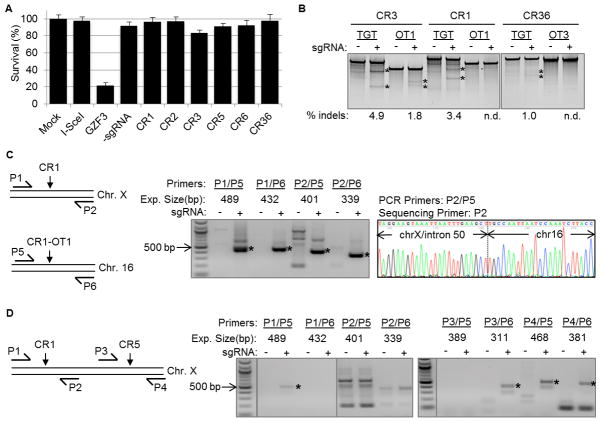
The CRISPR/Cas9 genome-editing platform is a promising technology to correct the genetic basis of hereditary diseases. The versatility, efficiency and multiplexing capabilities of the CRISPR/Cas9 system enable a variety of otherwise challenging gene correction strategies. Here, we use the CRISPR/Cas9 system to restore the expression of the dystrophin gene in cells carrying dystrophin mutations that cause Duchenne muscular dystrophy (DMD). We design single or multiplexed sgRNAs to restore the dystrophin reading frame by targeting the mutational hotspot at exons 45-55 and introducing shifts within exons or deleting one or more exons. Following gene editing in DMD patient myoblasts, dystrophin expression is restored in vitro. Human dystrophin is also detected in vivo after transplantation of genetically corrected patient cells into immunodeficient mice. Importantly, the unique multiplex gene-editing capabilities of the CRISPR/Cas9 system facilitate the generation of a single large deletion that can correct up to 62% of DMD mutations.


Conflict of interest statement

Conflict of interest statement: C.A.G. and D.G.O. have filed patent applications related to genome editing for Duchenne muscular dystrophy. C.A.G. is a scientific advisor to Editas Medicine, a company engaged in therapeutic development of genome editing technologies.

Figures

Figure 1
Figure 1. CRISPR/Cas9 targeting of the dystrophin gene
(A) sgRNA sequences were designed to bind sequences in the exon 45–55 mutational hotspot region of the dystrophin gene, such that gene editing could restore dystrophin expression from a wide variety of patient-specific mutations. Arrows within introns indicate sgRNA targets designed to delete entire exons from the genome. Arrows within exons indicate sgRNA targets designed to create targeted frameshifts in the dystrophin gene. (B) Example of frame correction following introduction of small insertions or deletions by NHEJ DNA repair in exon 51 using the CR3 sgRNA. (C) Schematic of multiplex sgRNA targets designed to delete exon 51 and restore the dystrophin reading frame in a patient mutation with the deletion of exons 48–50. (D) Schematic of multiplex sgRNA targets designed to delete the entire exon 45–55 region to address a variety of DMD patient mutations.
Figure 2
Figure 2. Fluorescence-activated flow sorting to enrich genetically modified DMD myoblasts
(A) A plasmid expressing a human-codon optimized SpCas9 protein linked to a GFP marker using a T2A ribosomal skipping peptide sequence was co-electroporated into human DMD myoblasts with one or two plasmids carrying sgRNA expression cassettes. (B) The indicated sgRNA expression cassettes were independently co-transfected into HEK293Ts with a separate plasmid expressing SpCas9 with (bottom) or without (top) a GFP marker linked to SpCas9 by a T2A ribosomal skipping peptide sequence. Gene modification frequencies were assessed at 3 days post-transfection by the Surveyor assay. (C) DMD myoblasts with deletions of exons 48–50 in the dystrophin gene were treated with sgRNAs that correct the dystrophin reading frame in these patient cells. Gene modification was assessed at 20 days post-electroporation in unsorted (bulk) or GFP+ sorted cells. (D) GFP expression in DMD myoblasts 3 days after electroporation with indicated expression plasmids. Transfection efficiencies and sorted cell populations are indicated by the gated region.
Figure 3
Figure 3. Targeted frameshifts to restore the dystrophin reading frame using CRISPR/Cas9
(A) The 5′ region of exon 51 was targeted using a sgRNA, CR3, that binds immediately upstream of the first out-of-frame stop codon. PAM: protospacer-adjacent motif. (B) The exon 51 locus was PCR amplified from HEK293T cells treated with SpCas9 and CR3 expression cassettes. Sequences of individual clones were determined by Sanger sequencing. The top sequence (bolded, exon in red) is the native, unmodified sequence. The number of clones for each sequence is indicated in parentheses. (C) Summary of total gene editing efficiency and reading frame conversions resulting from gene modification shown in (B). (D) Western blot for dystrophin expression in human DMD myoblasts treated with SpCas9 and the CR3 sgRNA expression cassette (Figure 2C) to create targeted frameshifts to restore the dystrophin reading frame. Dystrophin expression was probed using an antibody against the rod-domain of the dystrophin protein after 6 days of differentation.
Figure 4
Figure 4. Deletion of exon 51 from the human genome using multiplex CRISPR/Cas9 gene editing
(A) End-point genomic PCR across the exon 51 locus in human DMD myoblasts with a deletion of exons 48–50. The top arrow indicates the expected position of full-length PCR amplicons and the two lower arrows indicate the expected position of PCR amplicons with deletions caused by the indicated sgRNA combinations. (B) PCR products from (A) were cloned and individual clones were sequenced to determine insertions and deletions present at the targeted locus. The top row shows the wild-type unmodified sequence and the triangles indicate SpCas9 cleavage sites. At the right are representative chromatograms showing the sequences of the expected deletion junctions. (C) End-point RT-PCR analysis of dystrophin mRNA transcripts in CRISPR/Cas9-modified human Δ48–50 DMD myoblasts treated with the indicated sgRNAs. A representative chromatogram of the expected deletion PCR product is shown at the right. Asterisk: band resulting from hybridization of the deletion product strand to the unmodified strand. (D) Rescue of dystrophin protein expression by CRISPR/Cas9 genome editing was assessed by western blot for the dystrophin protein with GAPDH as a loading control. The arrow indicates the expected restored dystrophin protein band.
Figure 5
Figure 5. Deletion of exon 45–55 region in human DMD myoblasts by multiplex CRISPR/Cas9 gene editing
(A) End-point genomic PCR of genomic DNA to detect deletion of the region between intron 44 and intron 55 after treating HEK293Ts or DMD myoblasts with the indicated sgRNAs. (B) Individual clones of PCR products of the expected size for the deletions from DMD myoblasts in (A) were analyzed by Sanger sequencing to determine the sequences of genomic deletions present at the targeted locus. Below is a representative chromatograms showing the sequence of the expected deletion junctions. (C) End-point RT-PCR analysis of dystrophin mRNA transcripts in CRISPR/Cas9-modified human Δ48–50 DMD myoblasts treated with the indicated sgRNAs. A representative chromatogram of the expected deletion PCR product is shown at the right. (D) Analysis of restored dystrophin protein expression by western blot following electroporation of DMD myoblasts with sgRNAs targeted to intron 44 and/or intron 55.
Figure 6
Figure 6. Expression of restored human dystrophin in vivo following cell transplantation
Human Δ48–50 DMD myoblasts were treated with SpCas9, CR1, and CR5 to delete exon 51 and sorted for GFP expression as shown in Figure 2. These sorted cells and untreated control cells were injected into the hind limbs of immunodeficient mice and assessed for human-specific protein expression in muscle fibers after 4 weeks post-transplantation. Cryosections were stained with anti-human spectrin, which is expressed by both uncorrected and corrected myoblasts that have fused into mouse myofibers, or anti-human dystrophin antibodies as indicated. White arrows indicate muscle fibers positive for human dystrophin. Scale bars indicate 100 μm.
Figure 7
Figure 7. Evaluation of CRISPR/Cas9 toxicity and off-target effects for deletion of human exon 51
(A) Results of a cytotoxicity assay in HEK293T cells treated with human-optimized SpCas9 and the indicated sgRNA constructs. Cytotoxicity is based on survival of GFP-positive cells that are co-transfected with the indicated nuclease. I-SceI is a well-characterized non-toxic meganuclease and GZF3 is a known toxic zinc finger nuclease. n=3 independent transfections (mean + s.e.m.). (B) Surveyor analysis at off-target sites in sorted hDMD cells treated with expression cassettes encoding Cas9 the indicated sgRNAs. These three off-target sites tested in hDMD cells were identified from a panel of 50 predicted sites tested in HEK293T cells (Supplementary Figure 6 and Supplementary Table 2). TGT: on-target locus for indicated sgRNA. OT:off-target locus. (C, D) End-point nested PCR to detect chromosomal translocations in (C) HEK293T cells treated with Cas9 and CR1 or (D) sorted hDMD cells treated with Cas9, CR1, and CR5. The schematic depicts the relative location of nested primer pairs customized for each translocation event. The expected size of each band was estimated based on the primer size and the location of the predicted sgRNA cut site at each locus. Asterisks indicate bands detected at the expected size. The identities of the bands in (C) were verified by Sanger sequencing from each end (Supplementary Figure 11). A representative chromatogram for the P2/P5 translocation in HEK293T cells is shown.

Comment in

Similar articles

Cited by

References

    1. Gaj T, Gersbach CA, Barbas CF., 3rd ZFN, TALEN, and CRISPR/Cas-based methods for genome engineering. Trends Biotechnol. 2013;31:397–405. - PMC - PubMed
    1. Joung JK, Sander JD. TALENs: a widely applicable technology for targeted genome editing. Nat Rev Mol Cell Biol. 2013;14:49–55. - PMC - PubMed
    1. Silva G, Poirot L, Galetto R, Smith J, Montoya G, Duchateau P, et al. Meganucleases and other tools for targeted genome engineering: perspectives and challenges for gene therapy. Current gene therapy. 2011;11:11–27. - PMC - PubMed
    1. Sander JD, Joung JK. CRISPR-Cas systems for editing, regulating and targeting genomes. Nat Biotechnol. 2014;32:347–355. - PMC - PubMed
    1. Mali P, Yang L, Esvelt KM, Aach J, Guell M, DiCarlo JE, et al. RNA-guided human genome engineering via Cas9. Science. 2013;339:823–826. - PMC - PubMed

Publication types

Associated data