Review
. 2015;16(1):31-48.
doi: 10.2174/1389203716666150213160438.

Glucosidase II and MRH-domain containing proteins in the secretory pathway

Affiliations
Review

Glucosidase II and MRH-domain containing proteins in the secretory pathway

Cecilia D'Alessio et al. Curr Protein Pept Sci. 2015.

Abstract

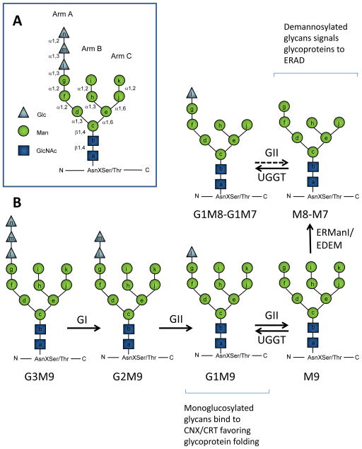
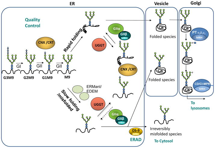
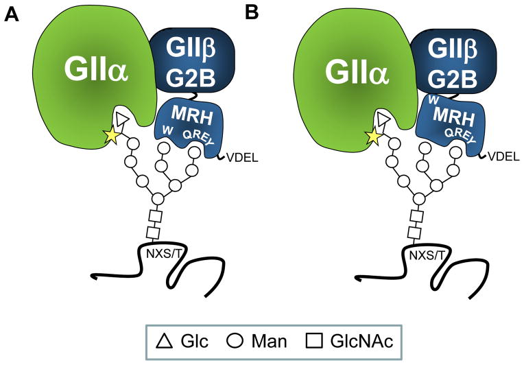
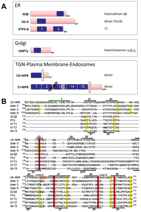
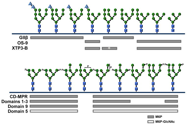
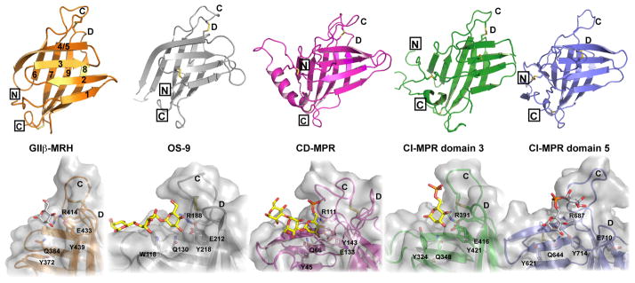
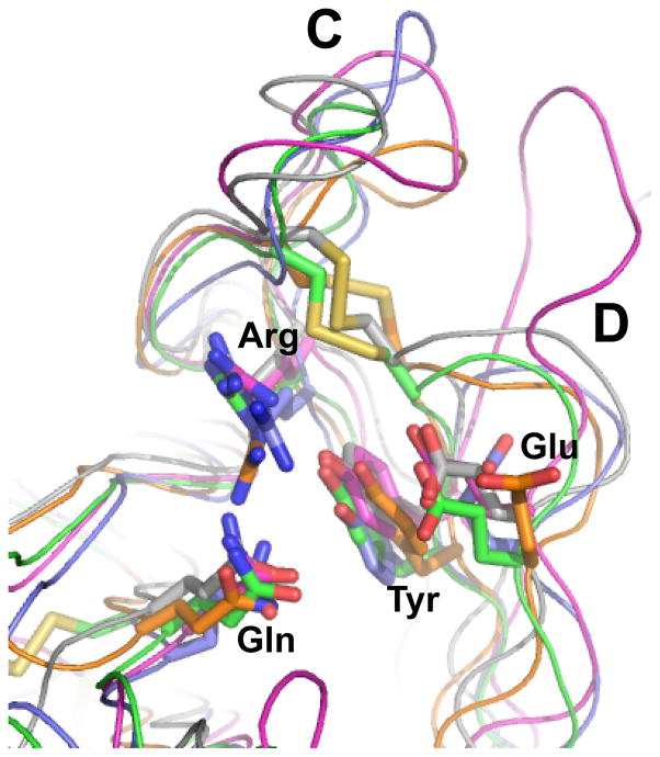
N-glycosylation in the endoplasmic reticulum (ER) consists of the transfer of a preassembled glycan conserved among species (Glc3Man9GlcNAc2) from a lipid donor to a consensus sequence within a nascent protein that is entering the ER. The protein-linked glycans are then processed by glycosidases and glycosyltransferases in the ER producing specific structures that serve as signalling molecules for the fate of the folding glycoprotein: to stay in the ER during the folding process, to be retrotranslocated to the cytosol for proteasomal degradation if irreversibly misfolded, or to pursue transit through the secretory pathway as a mature glycoprotein. In the ER, each glycan signalling structure is recognized by a specific lectin. A domain similar to that of the mannose 6-phosphate receptors (MPRs) has been identified in several proteins of the secretory pathway. These include the beta subunit of glucosidase II (GII), a key enzyme in the early processing of the transferred glycan that removes middle and innermost glucoses and is involved in quality control of glycoprotein folding in the ER (QC), the lectins OS-9 and XTP3-B, proteins involved in the delivery of ER misfolded proteins to degradation (ERAD), the gamma subunit of the Golgi GlcNAc-1-phosphotransferase, an enzyme involved in generating the mannose 6-phosphate (M6P) signal for sorting acidic hydrolases to lysosomes, and finally the MPRs that deliver those hydrolytic enzymes to the lysosome. Each of the MRH-containing proteins recognizes a different signalling N-glycan structure. Three-dimensional structures of some of the MRH domains have been solved, providing the basis to understand recognition mechanisms.


Conflict of interest statement

CONFLICT OF INTERESTS. None declared

Figures

Figure 1
Figure 1. Processing of N-glycans in the ER
A. Oligosaccharide Glc3Man9GlcNAc2 (G3M9) is transferred to Asn residues on nascent polypeptides by oligosaccharyltransferase. Lettering a–n indicates the order of addition of the monosaccharides during in vivo synthesis of the Dolichol-PP-Glc3Man9GlcNAc2 precursor. Arm A, B and C indicate the oligosaccharide branch. During biosynthesis residues a–g are added on the cytosolic face of the ER membrane from nucleotide-sugar precursors, while residues h–n are added from Dol-P-Glc or Dol-P-Man precursors after the oligosaccharide has flipped across the membrane. B. After glycan transfer to proteins, Glucosidase I (GI) removes glucose n, Glucosidase II (GII) removes glucose m and l, and UDP-Glc:glycoprotein glucosyltransferase (UGGT) adds glucose l. ER mannosidases may remove mannoseI and k. Monoglucosylated N-glycans are able to interact with ER lectins calnexin (CNX) and/or calreticulin (CRT). ER mannosidase I (ERManI) (MNSI in yeasts) acts as a timer of permanence of glycoproteins in the ER and removes residue I generating M8B. Subsequently, ERManI or other ER mannosidases (probably EDEM in mammals or Htm1in yeasts) may remove residue k, generating M7. Demannosylated glycans constitute the ERAD signal. Dotted lines are used to indicate that GII activity is reduced toward demmanosylated species.
Figure 2
Figure 2. Quality control of glycoprotein folding in the endoplasmic reticulum, ERAD and sorting in the secretory pathway
The glycan G3M9 transferred to proteins during N-glycosylation is immediately trimmed by glucosidases GI and GII. Monoglucosylated species generated by GII may interact with lectin/chaperones CNX or CRT, thus facilitating folding, preventing aggregation and providing a mechanism for ER retention of misfolded species. A second cleavage by GII liberates glycoproteins from the CNX/CRT anchor. This is a check point in the secretory pathway: if the proteins have acquired their native conformation, they can continue to transit through the secretory pathway to their final destination. If not yet properly folded, UGGT adds a glucose unit to allow another round of interactions between misfolded glycoproteins and lectin/chaperones. GII is also responsible for the removal of the glucose added by UGGT. Cycles of deglucosylation and reglucosylation catalyzed by the opposing activities of UGGT and GII continue until the glycoproteins acquire their native tertiary structure, thereby allowing their transit to their final destination. Misfolded/slow-folding species are characterized by ER mannosidase(s) (ERManI/EDEM)-catalyzed N-glycan demannosylation. OS-9 recognizes Manα 1,6Man on the trimmed C arm and facilitates entry of misfolded glycoproteins into the ERAD pathway where the misfolded glycoproteins exit the ER and are degraded by the proteosome in the cytosol. A decrease in N-glycan mannose content significantly diminishes in vivo GII-mediated deglucosylation rates but does not affect in vivo UGGT-mediated glucosylation, thus increasing the possibility of displaying monoglucosylated structures able to interact with CNX/CRT for longer time periods, and providing one more chance to escape from ERAD. If the final destination of a glycoprotein is the lysosome (as for acidic hydrolases), a M6P tag is added by UDP-N-acetylglucosamine:lysosomal enzyme GlcNAc-1-phosphotranfserase (PT) and the N-acetylglucosamine-1-phosphodiester α-N-acetylglucosaminidase (UCE) in the Golgi. M6P receptors (CD-MPR and CI-MPR) recognize this tag and concentrate these proteins in clatrin-coated vesicles that bud from trans Golgi network. MRH domain-containing proteins present in GIIβ subunit, OS-9, PT γ subunit and CD-MPR or CI-MPR are indicated.
Figure 3
Figure 3. Proposed models for GIIβ MRH domain-mediated enhancement of N-glycan deglucosylation
GII is a heterodimer composed of a GIIα catalytic and a GIIβ regulatory subunit. GIIβ enhances GII deglucosylation activity towards both G2M9 and G1M9 through its MRH domain. Upon binding mannose units in the B and/or C arms of the glycan, the GIIβ MRH domain presents bonds to be cleaved to the GIIα catalytic site (star). GIIβ also provides the retention/retrieval signal for proper ER localization of the heterodimer (−ValAspGluLeu (VDEL) in S. pombe). G2B domain is involved in GIIα-GIIβ interaction. Residues Gln-384, Arg-414, Glu-433, Tyr-439 (QREY) form the binding pocket that is the “signature motif” for MRH domain-containing proteins. There are two possible models for the role of Trp-409 in GII activity: In (A) mannose-binding essential residues Gln-384, Arg-414, Glu-433, Tyr-439 form a pocket which binds arm C of the glycan, while residue Trp-409 (W) interacts with arm B. This bidentate interaction allows the glucose-containing arm A to be juxtaposed to GIIα’s catalytic site. In (B) Trp-409 interacts with other regions of the β-subunit and influences its affinity for N-glycans. These models suggest that removal of mannoses by ER mannosidases will reduce both the binding of the glycan and GII activity.
Figure 4
Figure 4. A) Schematic diagram of human MRH domain-containing proteins
The location of the MRH domains (blue) is shown. The CI-MPR contains 15 contiguous MRH domains, with MRH domain 13 containing a 48-residue fibronectin type II (FnII) insert (gray). The CD-MPR and CI-MPR are type I integral membrane proteins and the location of the single transmembrane domain is shown by a vertical hatched bar. The number of amino acids in each protein, including the N-terminal signal sequence that is not shown, is indicated. The oligomeric state of the protein is listed (note: the oligomeric state of human OS-9 has not been established). GIIβ is the non-catalytic subunit of the heterodimeric glycosidase, glucosidase II, a resident protein of the ER. OS-9 and XTP3-B are also resident proteins of the ER. The γ-subunit of GlcNAc-1-phosphotransferase is the non-catalytic subunit of this hexameric glycosyltransferase localized to early Golgi compartments. CD-MPR and CI-MPR constitutively recycle between TGN, endosomes and plasma membrane. ER = endoplasmic reticulum, TGN = trans Golgi network. B) Structure-based sequence alignment. Structure-based sequence alignment of the MRH domains of bovine CD-MPR (A27068), domains 3, 5 and 9 of the bovine CI-MPR (A30788), human GII β subunit (CAA04006), human GlcNAc-1-phosphotransferase γ subunit (Q9UJJ9), the N- and C-terminal MRH domains of human XTP3-B (NP_056516), and human OS-9 (BAA24363). GII β subunit, OS-9, XTP3-B, and GlcNAc-1-phosphotransferase γ subunit contain all four essential residues for M6P recognition (Gln, Arg, Glu, and Tyr), which are shaded in red. The WW motif of OS-9 is boxed in orange. Y679, which is present in the binding pocket of domain 5, is boxed in blue. The residues known to bind the phosphate group in the CD-MPR (D103, N104, H105) and CI-MPR MRH domain 3 (S386) are boxed in red. The conserved tryptophan residue in loop C of GII β subunit is boxed in green. Residues in OS-9 predicted to prevent binding of the phosphate moiety of M6P (D182, L183) are boxed in green. The cysteine residues are shaded in yellow. The secondary structural elements of CD-MPR and OS-9 are shown, with dark blue arrows representing the β-strands and the green line representing an α-helix. Location of loops C and D are shown. PTγ = GlcNAc-1-phosphotransferase γ subunit, Erl1 = XTP3-B N-terminal MRH domain, Erl2 = XTP3-B C-terminal MRH domain
Figure 5
Figure 5. Summary of glycan specificity of the MRH domain-containing proteins
Top panel, Binding of glucosidase II β-subunit (GIIβ), OS-9, and XTP3-B to high mannose-type glycans is shown. The glycan binding specificity of XTP3-B remains unresolved, with recent studies indicating a preference for M9 glycan (asterisk-labeled bar) wheras other groups report binding to M5, M6 and M7 glycans. GlcNAc-1-phosphotransferase γ subunit’s ability to interact with specific glycan structures has not yet been determined. Bottom panel, Binding of MPRs to phosphorylated glycans. The CD-MPR and each of the three carbohydate binding domains of the CI-MPR recognize different populations of phosphorylated glycans. For simplicity, only the M6P (P) containing glycans are shown, with the phosphorylated mannose residue highlighted. M6P-GlcNAc-containing glycans have the identical structure, except with a phosphodiester at the analogous position as the phosphomonoester. Domains 1–3 and 9 preferentially bind M6P-containing glycans (grey bars), whereas domain 5 binds only M6P-GlcNAc-containing glycans (open bar). Both MPRs are unable to bind a M7 glycan containing a phosphomannosyl residue on the B (α1,3 linked mannose h) or C (α1,6 linked mannose j) arm. Residues are labeled as in Figure 1A: glucose (blue triangles), mannose (green circles) and GlcNAc (blue squares).
Figure 6
Figure 6. Comparison of the structures of the MRH domains
Top, Ribbon diagram of the MRH domains of S. pombe GIIβ (orange, solution structure, PDB ID: 2LVX), human OS-9 (gray, crystal structure, PDB ID: 3AIH), bovine CD-MPR (magenta, crystal structure, PDB ID: 1C39), bovine CI-MPR MRH domain 3 (green, crystal structure, PDB ID: 1SZO), bovine CI-MPR MRH domain 5 (blue, solution structure, PDB ID: 2KVB). Disulfide bridges are shown in yellow, N and C termini are boxed, β-strands are numbered from the N to C terminus, and loops C and D are labeled. Bottom, Close-up view of the carbohydrate binding sites are shown below the respective ribbon diagram. The four essential residues for mannose binding are shown along with the proposed linkage-sensing Tyr or Trp. Molecular surfaces are shown in gray over the ribbon diagram. Carbohydrate ligands are depicted as ball-and-stick. Structures solved in the presence of a bound ligand (Manα1,6Manα1,6Man for OS-9, pentamannosyl phosphate for CD-MPR, M6P for CI-MPR MRH domain 3) are shown in yellow. Modeled ligands are depicted in gray (mannose for GIIβ, methyl-M6P-GlcNAc for CI-MPR MRH domain 5) and are placed in the binding pocket based on superimposition of the four essential residues with a known MRH domain structure containing a bound ligand.
Figure 7
Figure 7. Close-up view of the carbohydrate binding pocket
Overlay of the four essential residues (Gln, Arg, Glu, Tyr) of S. pombe GIIβ (orange, PDB ID: 2LVX), human OS-9 (gray, PDB ID: 3AIH), bovine CD-MPR (magenta, PDB ID: 1C39), bovine CI-MPR MRH domain 3 (green, PDB ID: 1SZO), bovine CI-MPR MRH domain 5 (blue, PDB ID: 2KVB). Disulfide bridges are shown in yellow and loops C and D are labeled.

References

    1. D’Alessio C, Caramelo JJ, Parodi AJ. UDP-Glc:Glycoprotein glucosyltransferase-glucosidase II, the ying-yang of the er quality control. Semin Cell Dev Biol. 2010;21(5):491–9. - PMC - PubMed
    1. Kornfeld R, Kornfeld S. Assembly of asparagine-linked oligosaccharides. Annual Review of Biochemistry. 1985;54:631–64. - PubMed
    1. Aebi M, Bernasconi R, Clerc S, Molinari M. N-glycan structures: Recognition and processing in the er. Trends Biochem Sci. 2010;35(2):74–82. - PubMed
    1. Apweiler R, Hermjakob H, Sharon N. On the frequency of protein glycosylation, as deduced from analysis of the swiss-prot database. Biochim Biophys Acta. 1999;1473(1):4–8. - PubMed
    1. Nilsson IM, von Heijne G. Determination of the distance between the oligosaccharyltransferase active site and the endoplasmic reticulum membrane. J Biol Chem. 1993;268(8):5798–801. - PubMed

Publication types

MeSH terms