. 2015 Feb 18;10(2):e0115991.
doi: 10.1371/journal.pone.0115991. eCollection 2015.

Cardiac telocytes and fibroblasts in primary culture: different morphologies and immunophenotypes

Affiliations

Cardiac telocytes and fibroblasts in primary culture: different morphologies and immunophenotypes

Yihua Bei et al. PLoS One. .

Abstract

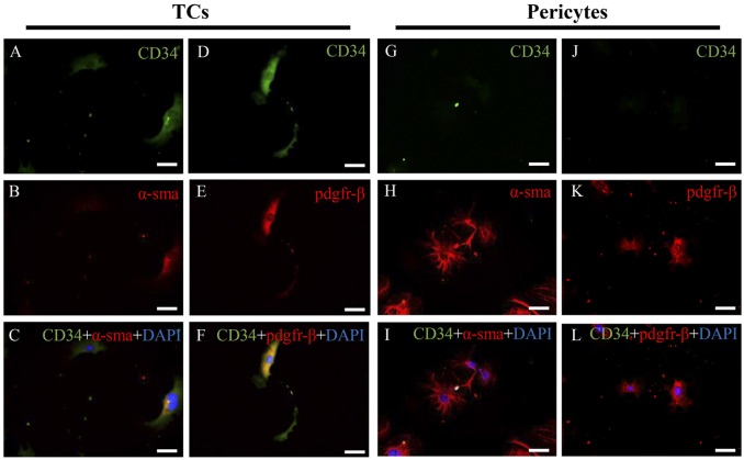
Telocytes (TCs) are a peculiar type of interstitial cells with very long prolongations termed telopodes. TCs have previously been identified in different anatomic structures of the heart, and have also been isolated and cultured from heart tissues in vitro. TCs and fibroblasts, both located in the interstitial spaces of the heart, have different morphologies and functionality. However, other than microscopic observation, a reliable means to make differential diagnosis of cardiac TCs from fibroblasts remains unclear. In the present study, we isolated and cultured cardiac TCs and fibroblasts from heart tissues, and observed their different morphological features and immunophenotypes in primary culture. Morphologically, TCs had extremely long and thin telopodes with moniliform aspect, stretched away from cell bodies, while cell processes of fibroblasts were short, thick and cone shaped. Furthermore, cardiac TCs were positive for CD34/c-kit, CD34/vimentin, and CD34/PDGFR-β, while fibroblasts were only vimentin and PDGFR-β positive. In addition, TCs were also different from pericytes as TCs were CD34 positive and α-SMA weak positive while pericytes were CD34 negative but α-SMA positive. Besides that, we also showed cardiac TCs were homogenously positive for mesenchymal marker CD29 but negative for hematopoietic marker CD45, indicating that TCs could be a source of cardiac mesenchymal cells. The differences in morphological features and immunophenotypes between TCs and fibroblasts will provide more compelling evidence to differentiate cardiac TCs from fibroblasts.


Conflict of interest statement

Competing Interests: The authors have declared that no competing interests exist.

Figures

Fig 1
Fig 1. Light microscope shows cardiac telocytes (TCs, A-F) and fibroblasts (FB, G-L) in primary culture from heart tissues at 48 h, 72 h, and 96 h after the day of primary culture.
Arrows show typical TCs with long and thin telopodes in primary culture. Original magnification 200×; Scale bar = 50 μm.
Fig 2
Fig 2. Light microscope shows telocytes (TCs) with typical morphological features.
(A) A TC with small cell body and extremely thin and long telopode (Tp). (B) and (C) A TC with significant moniliform aspect: alternation of podoms-podomeres (enlarged in inset). (D-F) show more TCs with typical morphological features. Original magnification 200×; Scale bar = 50 μm.
Fig 3
Fig 3. Double immunofluorescent staining for CD34/c-kit, CD34/vimentin and CD34/PDGFR-β of cardiac telocytes (TCs) and fibroblasts (FB) in primary culture.
Fluorescent inverted microscope shows that TCs are positive for (A) CD34 (green) and (B) c-kit (red), while FBs are negative for (J) CD34 (green) and (K) c-kit (red). Co-localization of c-kit and CD34 (yellow) is significant in TCs (C), while absent in FBs (L). TCs are positive for (D) CD34 (green) and (E) vimentin (red). FBs are negative for (M) CD34 (green), but positive for (N) vimentin (red). Co-localization of vimentin and CD34 (yellow) is significant in TCs (F), while absent in FBs (O). TCs are positive for (G) CD34 (green) and (H) PDGFR-β (red). FBs are negative for (P) CD34 (green), but positive for (Q) PDGFR-β (red). Co-localization of PDGFR-β and CD34 (yellow) is significant in TCs (I), while absent in FBs (R). Nuclei are counterstained with DAPI (blue). Original magnification 200×; Scale bar = 50 μm. BF, bright field.
Fig 4
Fig 4. Double immunofluorescent staining for CD34/α-SMA and CD34/PDGFR-β of cardiac telocytes (TCs) and pericytes in primary culture.
Fluorescent inverted microscope shows that TCs are positive for (A) CD34 (green) and relatively weak positive for (B) α-SMA (red), while pericytes are negative for (G) CD34 (green) but strong positive for (H) PDGFR-β (red). Co-localization of α-SMA and CD34 (yellow) is extremely weak in TCs (C), while absent in pericytes (I). TCs are positive for (D) CD34 (green) and (E) PDGFR-β (red), while pericytes are negative for (J) CD34 (green), but positive for (K) PDGFR-β (red). Co-localization of PDGFR-β and CD34 (yellow) was significant in TCs (F), while absent in pericytes (L). Nuclei were counterstained with DAPI (blue). Original magnification 200×; Scale bar = 50 μm. BF, bright field.
Fig 5
Fig 5. Cell surface markers of cardiac telocytes (TCs) and bone marrow-derived mesenchymal stem cells (BMSC).
Cells collected are shown in A and D. Flow cytometry analysis show that cardiac TCs are homogenously positive for mesenchymal marker CD 29 (B) as BMSC (E), but negative for hematopoietic marker CD45 (C) as BMSC (F). Unlabeled cell (black) controls are included for comparison. FSC: forward scatter; SSC,side scatter.

References

    1. Popescu LM, Faussone-Pellegrini M-S (2010) TELOCYTES—a case of serendipity: the winding way from Interstitial Cells of Cajal (ICC), via Interstitial Cajal-Like Cells (ICLC) to TELOCYTES. J Cell Mol Med 14:729–740. 10.1111/j.1582-4934.2010.01059.x - DOI - PMC - PubMed
    1. Popescu LM, Manole CG, Gherghiceanu M, Ardelean A, Nicolescu MI, et al. (2010) Telocytes in human epicardium. J Cell Mol Med 14:2085–2093. 10.1111/j.1582-4934.2010.01129.x - DOI - PMC - PubMed
    1. Gherghiceanu M, Manole CG, Popescu LM (2010) Telocytes in endocardium: electron microscope evidence. J Cell Mol Med 14:2330–2334. 10.1111/j.1582-4934.2010.01133.x - DOI - PMC - PubMed
    1. Hinescu ME, Gherghiceanu M, Suciu L, Popescu LM (2011) Telocytes in pleura: two- and three-dimensional imaging by transmission electron microscopy. Cell Tissue Res 343:389–397. 10.1007/s00441-010-1095-0 - DOI - PMC - PubMed
    1. Rusu MC, Jianu AM, Mirancea N, Didilescu AC, Mănoiu VS, et al. (2012) Tracheal telocytes. J Cell Mol Med 16:401–405. 10.1111/j.1582-4934.2011.01465.x - DOI - PMC - PubMed

Publication types