. 2015 Feb 18;11(2):e1004699.
doi: 10.1371/journal.ppat.1004699. eCollection 2015 Feb.

Direct binding of retromer to human papillomavirus type 16 minor capsid protein L2 mediates endosome exit during viral infection

Affiliations

Direct binding of retromer to human papillomavirus type 16 minor capsid protein L2 mediates endosome exit during viral infection

Andreea Popa et al. PLoS Pathog. .

Abstract

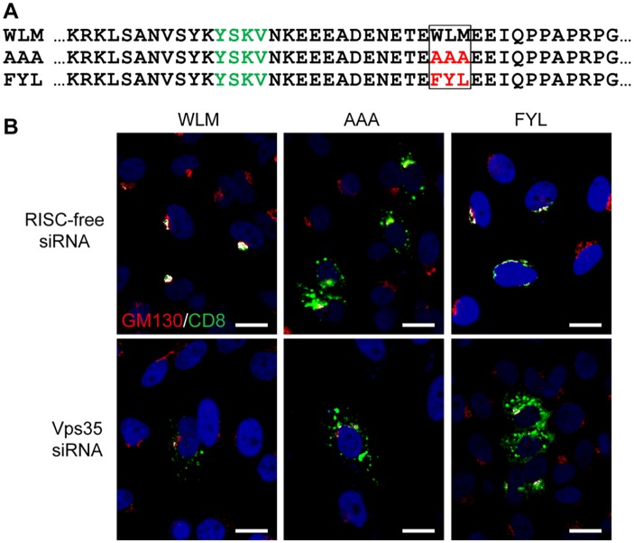
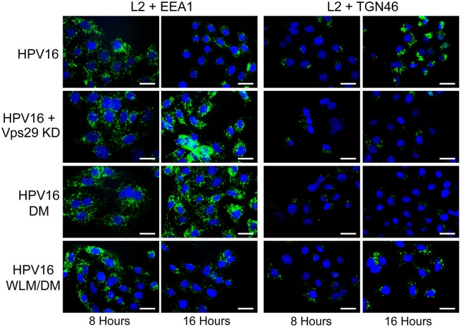
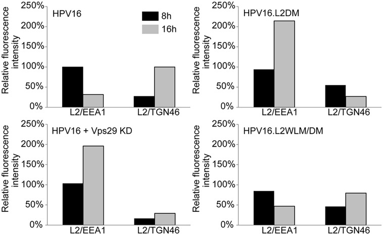
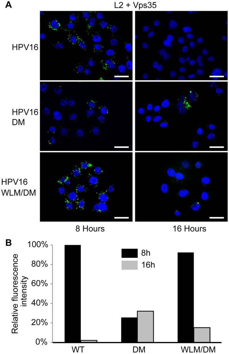
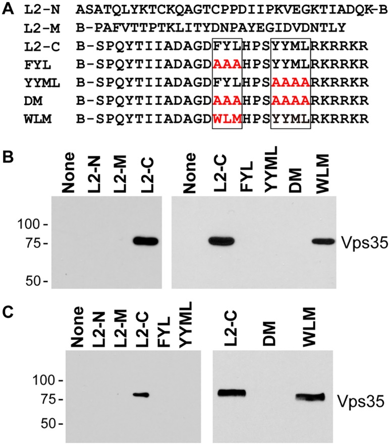
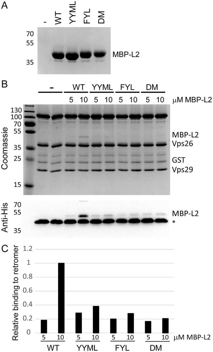
Trafficking of human papillomaviruses to the Golgi apparatus during virus entry requires retromer, an endosomal coat protein complex that mediates the vesicular transport of cellular transmembrane proteins from the endosome to the Golgi apparatus or the plasma membrane. Here we show that the HPV16 L2 minor capsid protein is a retromer cargo, even though L2 is not a transmembrane protein. We show that direct binding of retromer to a conserved sequence in the carboxy-terminus of L2 is required for exit of L2 from the early endosome and delivery to the trans-Golgi network during virus entry. This binding site is different from known retromer binding motifs and can be replaced by a sorting signal from a cellular retromer cargo. Thus, HPV16 is an unconventional particulate retromer cargo, and retromer binding initiates retrograde transport of viral components from the endosome to the trans-Golgi network during virus entry. We propose that the carboxy-terminal segment of L2 protein protrudes through the endosomal membrane and is accessed by retromer in the cytoplasm.


Conflict of interest statement

The authors have declared that no competing interests exist.

Figures

Fig 1
Fig 1. The carboxy-terminus of HPV16 L2 contains two potential retromer recognition motifs required for infectivity.
A. Alanine scanning mutagenesis of the carboxy-terminus of the HPV16 L2 protein. The top line shows amino acids 434 to 461 of the wild-type L2 protein, with the potential retromer binding motifs in boxes. Amino acid substitutions in the mutants are shown in red. The double mutant (DM) has both potential retromer binding sites replaced with alanines; WLM contains a known retromer signal in place of FYL but retains the wild-type YYML sequence; WLM/DM contains the WLM substitution and the YYML to AAAA substitution. B. HA-tagged PsVs containing equal numbers of the GFP reporter plasmid (corresponding to MOI of approximately 0.5 for the wild-type) were added to HeLa cells, and 48h post-infection GFP-positive cells were counted by flow cytometry. WT indicates wild-type PsV. Multiple independent pseudovirus stocks were tested for each mutant. Results shown are averages of four independent experiments, with error bars showing standard deviation. C. HeLa (black bars) and HaCaT cells (grey bars) were infected with equal numbers of FLAG-tagged PsV (corresponding to MOI of 0.5 in each cell type for the wild-type virus). Forty-eight hours post-infection, GFP-positive cells were counted by flow cytometry. WLM/DM, the FYL/AAA mutation in the double mutant was replaced with Trp-Leu-Met; WLM, FYL to WLM mutation only. Results shown are averages of multiple independent experiments with multiple independent PsV stocks. D. L2 sequences of HPV types 1 to 45 were retrieved from the Papillomavirus episteme PaVE database (http://pave.niaid.nih.gov/) and trimmed to a 26 amino acid segment centered on the L in FYL (position 13). Sequence conservation is shown in a Sequence Logo plot (http://weblogo.berkeley.edu/logo.cgi). Conserved FYL and related sequences are shown in box.
Fig 2
Fig 2. The L2 FYL sequence can act as a retromer sorting motif.
A. Sequences of the membrane-proximal cytoplasmic tail of CD8-CIMPR fusion proteins used in this experiment. The YSKV endocytosis motif in CIMPR is in green, mutations in the WLM retromer sorting signal are in red. B. HeLa-M cells were transfected with a plasmid expressing the wild-type CD8-CIMPR fusion protein (WLM) or a mutant fusion protein with mutations replacing WLM (AAA or FYL) and with control RISC-free siRNA or siRNA targeting retromer subunit Vps35, as indicated. After 24 hours, live cells were incubated with anti-CD8 for three hours, and localization of the construct was assessed by co-immunofluorescence with Golgi marker GM130. CD8-CIMPR fusion protein, green; GM130, red; nuclei, blue. A single plane in the Z-dimension is shown in each panel. Overlap between fusion protein and Golgi marker GM130 is pseudocolored white. The size bars on these and all subsequent images are 20 μM. (We note that HeLa-M cells have larger nuclei than the HeLa cells used in other experiments.) The component images used to generate this figure are shown in S4 Fig. Similar results were obtained in three independent experiments.
Fig 3
Fig 3. Retromer activity and potential retromer binding sites in L2 are required for exit from the early endosome and entry into the trans-Golgi network.
HeLa cells were transfected with siRNA targeting retromer subunit Vps29 (Vps29 KD) or left untreated. 48 hours later, cells were infected with FLAG-tagged wild-type HPV16, HPV16.L2DM or HPV16.L2WLM/DM PsV at MOI of 50 (according to reporter plasmid normalization). Eight or 16 hours post-infection, cells were fixed, permeabilized, and incubated with an anti-FLAG antibody and an antibody recognizing EEA1 (early endosome marker) or TGN46 (TGN marker). PLA was performed to assess L2 in proximity to the cellular marker (green). Nuclei are stained blue with DAPI. A single plane in the Z-dimension is shown in each panel. Similar results were obtained in multiple independent experiments with independent PsV stocks.
Fig 4
Fig 4. Quantitation of HPV trafficking in the absence of retromer activity or binding.
Images obtained as in Fig. 3 were processed by Blobfinder software to determine the PLA fluorescence intensity per cell in each sample. The average intensity for EEA1/L2 samples was normalized to the control sample infected with wild-type PsV at eight hours post-infection, and the average intensity for TGN46/L2 samples was normalized to the control sample infected with wild-type PsV at 16 hours post-infection. Black bars, eight hours; grey bars, 16 hours. The results show quantitation of a representative experiment. Because of the inherent variability of PLA assays, it was not possible to directly compare independent experiments, but similar results were obtained in two additional independent experiments.
Fig 5
Fig 5. Retromer sorting signals in L2 C-terminus are important for its association with retromer during entry.
A. HeLa cells were infected with FLAG-tagged HPV16, HPV16.L2DM or HPV16.L2WLM/DM PsV at MOI of 50. Eight or 16 hours post-infection, cells were fixed, permeabilized, and incubated with an anti-FLAG antibody and an antibody recognizing retromer subunit Vps35. PLA was performed to assess proximity of L2 and Vps35 (green). Nuclei are stained blue with DAPI. A single plane in the Z-dimension is shown in each panel. B. Images obtained as in panel A were processed by Blobfinder software to determine the PLA fluorescence intensity per cell in each sample. The average intensity was normalized to the control sample infected with wild-type PsV at eight hours post-infection. Black bars, eight hours; grey bars, 16 hours. The results show quantitation of a representative experiment. Similar results were obtained in two additional independent experiments.
Fig 6
Fig 6. Binding of carboxy-terminal L2 peptides to retromer.
A. The top lines show the sequences of biotinylated peptides, where B indicates position of biotin. Putative retromer recognition motifs in the carboxy-terminal peptide are shown in boxes. Mutant versions of the carboxy-terminal peptide are also shown, with the mutations in red. B. Left panel. L2-N, L2-M, or L2-C peptide was incubated with uninfected HeLa cell RIPA lysate. The samples were analyzed by streptavidin pull-down, SDS-PAGE, and immunoblotting with an anti-Vps35 antibody. Molecular weight markers in kDa are shown at the left. Right panel. The wild-type or a mutant carboxy-terminal peptide were incubated with uninfected HeLa cell HEPES lysate and processed as in panel A. Similar results were obtained in three or more independent experiments. C. Experiments performed as in panel B with HaCaT cell RIPA lysates.
Fig 7
Fig 7. L2 retromer motifs mediate direct binding to retromer.
A. MBP-L2 fusion proteins containing the wild-type or mutant carboxy-terminal segment of L2 were purified from E. coli and subjected to SDS-PAGE and staining with coomassie brilliant blue. Molecular weight markers are shown on the left. B. 5 or 10μM purified MBP-L2 fusion proteins were bound to purified retromer trimer, eluted, and subjected to SDS-polyacrylamine gel electrophoresis. Proteins were visualized by coomassie brilliant blue staining (top panel) or immunoblotting for the His-epitope on the MBP fusion protein (bottom panel). The band migrating at ~40 kDa indicated by the asterisks in all lanes in the immunoblot is Vps26, which cross-reacts with the anti-His antibody. Similar results were obtained in two independent experiments. C. The immunoblotting results in panel B were quantified and are plotted relative to binding of retromer to 10μM wild-type MBP-L2 fusion protein.

Similar articles

Cited by

References

    1. Parkin DM, Bray F (2006) Chapter 2: The burden of HPV-related cancers. Vaccine 24 Suppl 3: S3/11–25. - PubMed
    1. Liu WJ, Gissmann L, Sun XY, Kanjanahaluethai A, Muller M, et al. (1997) Sequence close to the N-terminus of L2 protein is displayed on the surface of bovine papillomavirus type 1 virions. Virology 227: 474–483. - PubMed
    1. Wang JW, Roden RB (2013) L2, the minor capsid protein of papillomavirus. Virology 445: 175–186. 10.1016/j.virol.2013.04.017 - DOI - PMC - PubMed
    1. Buck CB, Trus BL (2012) Chapter 18. The Papillomavirus Virion: A Machine Built to Hide Molecular Achilles’ Heels Viral Molecular Machines, Adv Exp Med Biol (Rossmann MG, Rao VB, eds) 726: 403–422. 10.1007/978-1-4614-0980-9_18 - DOI - PubMed
    1. Day PM, Lowy DR, Schiller JT (2008) Heparan sulfate-independent cell binding and infection with furin-precleaved papillomavirus capsids. J Virol 82: 12565–12568. 10.1128/JVI.01631-08 - DOI - PMC - PubMed

Publication types

MeSH terms