Comparative Study
. 2015 Oct;89(10):1811-25.
doi: 10.1007/s00204-015-1453-5. Epub 2015 Feb 19.

Diquat causes caspase-independent cell death in SH-SY5Y cells by production of ROS independently of mitochondria

Affiliations
Comparative Study

Diquat causes caspase-independent cell death in SH-SY5Y cells by production of ROS independently of mitochondria

R Nisar et al. Arch Toxicol. 2015 Oct.

Erratum in

Abstract

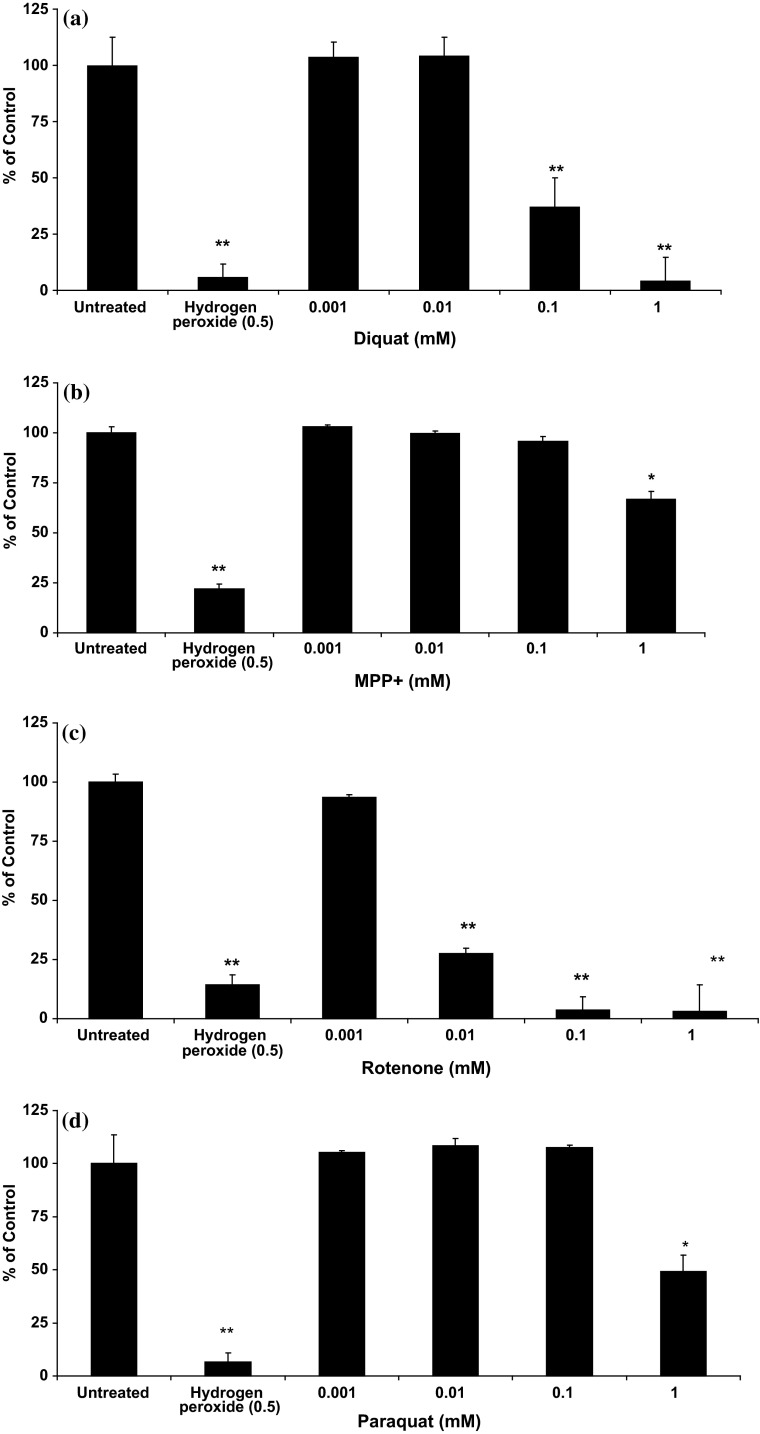
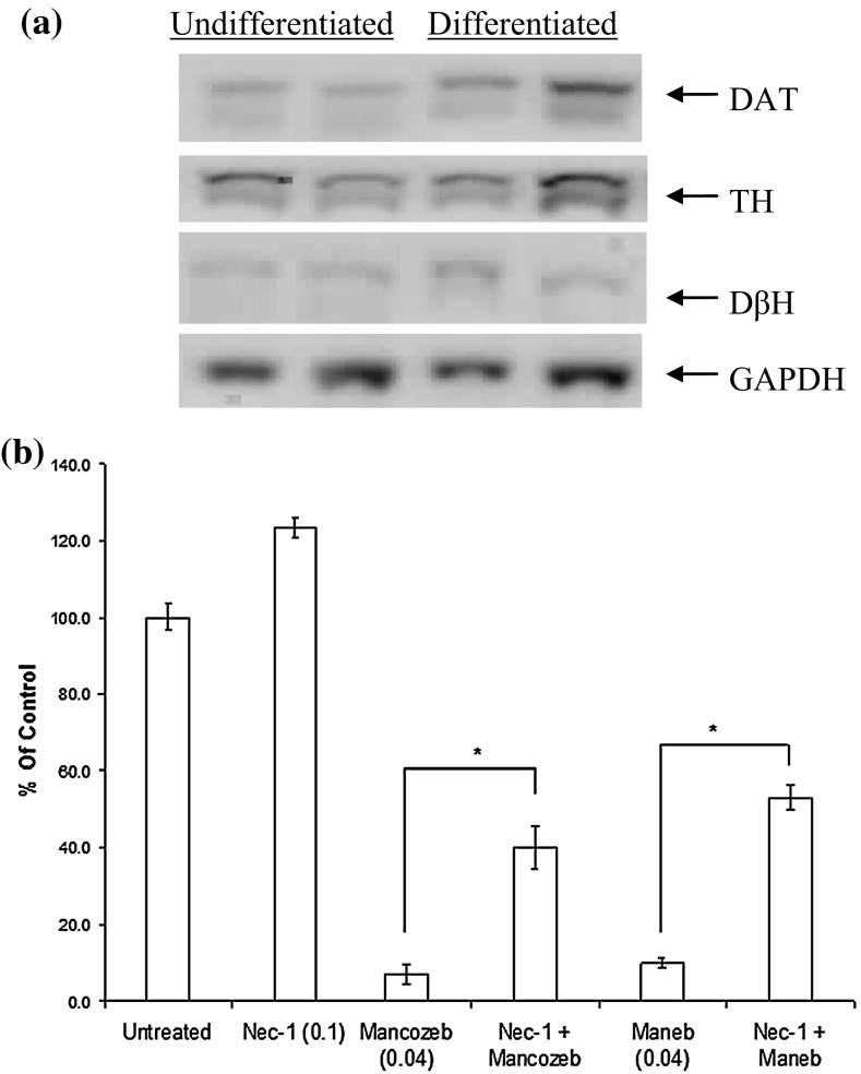
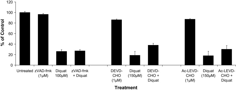
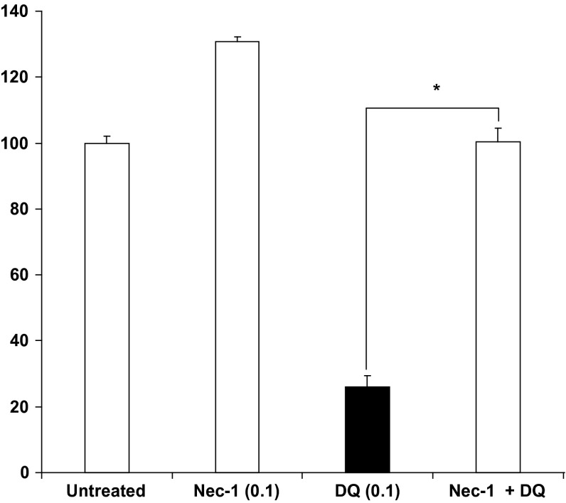
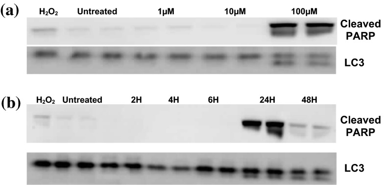
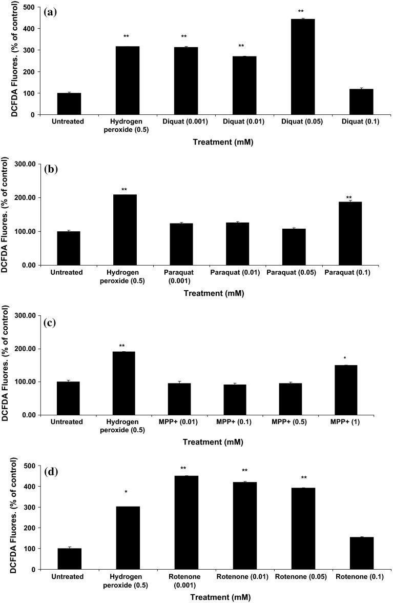
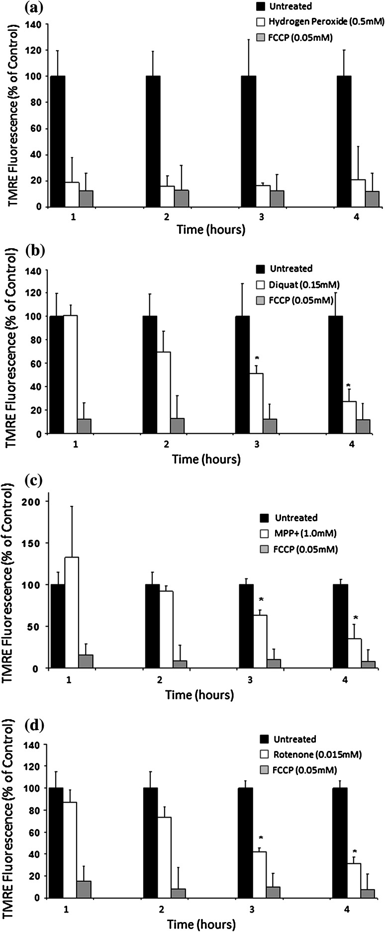
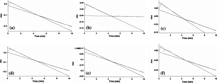
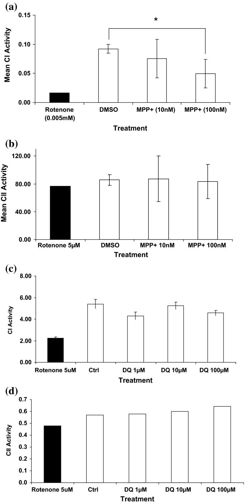
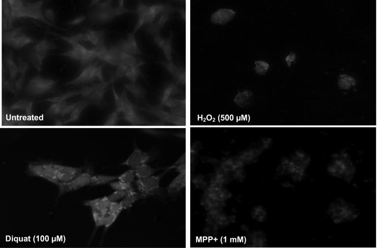
Evidence indicates that Parkinson's disease (PD), in addition to having a genetic aetiology, has an environmental component that contributes to disease onset and progression. The exact nature of any environmental agent contributing to PD is unknown in most cases. Given its similarity to paraquat, an agrochemical removed from registration in the EU for its suspected potential to cause PD, we have investigated the in vitro capacity of the related herbicide Diquat to cause PD-like cell death. Diquat showed greater toxicity towards SH-SY5Y neuroblastoma cells and human midbrain neural cells than paraquat and also MPTP, which was independent of dopamine transporter-mediated uptake. Diquat caused cell death independently of caspase activation, potentially via RIP1 kinase, with only a minor contribution from apoptosis, which was accompanied by enhanced reactive oxygen species production in the absence of major inhibition of complex I of the mitochondrial respiratory chain. No changes in α-synuclein expression were observed following 24-h or 4-week exposure. Diquat may, therefore, kill neural tissue by programmed necrosis rather than apoptosis, reflecting the pathological changes seen following high-level exposure, although its ability to promote PD is unclear.

Keywords: Apoptosis; Diquat; Mitochondria; Necrosis; Parkinson’s disease; Pesticide.


Figures

Fig. 1
Fig. 1
Effects of toxin treatment on SH-SYSY cell viability. Cells were incubated overnight with a range of toxin concentrations and cell viability determined using Alamar Blue reduction. a Diquat, b MPP+, c rotenone and d paraquat. Hydrogen peroxide (0.5 M) was used as a positive control. *P < 0.05, **P < 0.01, unpaired t test
Fig. 2
Fig. 2
a Western blot analyses of DAT in undifferentiated and differentiated SHSY5Y cells demonstrating the presence of dopamine transporter (DAT), tyrosine hydroxylase (TH) and dopamine β-hydroxylase (DβH) expression in both undifferentiated and differentiated cells after 5 days. b Effect of DAT inhibitors GBR 12909 and BTCP on cytotoxicity: cells were exposed to the dopamine transporter inhibitor GBR12909 for 2 h prior to exposure to diquat (DQ) with the continuous presence of DAT inhibitor. No significant reduction in toxicity was observed suggesting the DQ uptake into cells is not DAT mediated
Fig. 3
Fig. 3
Effect of caspase inhibition on cell viability. Cells were pre-incubated with zVAD.fmk (1 µM), DEVD-CHO (1 µM) or Ac-LEVD-CHO (1 µM) for 2 h before diquat addition. After 24-h incubation, cell viability was evaluated by Alamar Blue reduction assay. The data are expressed as mean ± SD (*P values <0.05 were accepted as significant). ns Not significant
Fig. 4
Fig. 4
Effects of necrostatin-1 on cell viability after diquat treatment. A cells were treated with 100 µM necrostatin-1 (Nec-1) for 2 h before addition of diquat (DQ) and continuously exposed to Nec-1 throughout. A significant increase in viability was observed following treatment (*P < 0.01)
Fig. 5
Fig. 5
Effect of diquat treatment on PARP-1 and LC3 expression. Expression of cleaved PARP-1 by Western blot analysis after a 24 h 1, 10 and 100 µM diquat exposure and b 48-h treatment at 100 µM diquat exposure with protein cleavage only occurring at 24-h exposure. Similar effects were seen with the expression of the autophagic marker protein LC3 – II6 (lower panel) after diquat exposure
Fig. 6
Fig. 6
Toxin-induced ROS production in SH-SY5Y cells. A dose-dependent increase in ROS production was noted with diquat (a), paraquat (b), MPP+ (c) and rotenone (d) (results are mean ± SD, of at least three replicates. P values <0.05* or <0.01** were accepted as significant)
Fig. 7
Fig. 7
Effect of diquat on mitochondrial trans membrane potential (∆Ψm). Cells were loaded with the mitochondrial redox sensitive dye TMRE and at selected time points following exposure TMRE fluorescence determined as a percentage of untreated cells. Hydrogen peroxide (a) showed a rapid loss of TMRE fluorescence similar to the mitochondrial uncoupler FCCP (0.001 mM). Diquat (0.150 mM, b), MPP+ (1 mM, c) and rotenone (0.0125 mM, d) showed a slower, time-dependant but significant reduction in TMRE fluorescence
Fig. 8
Fig. 8
Inhibition of NADH: quinone reductase (complex I) activity. Complex I activity (measured as rate of change of NADH oxidised in μmols of NADH oxidised/min) in a untreated (DMSO; without rotenone addition), b rotenone (5 µM), c diquat (40 µM), d paraquat (40 µM), e MPP+ (40 µM) and f MPTP (100 µM). Rotenone (5 µM) shows rapid and almost complete inhibition of activity, whilst other toxins show minimal inhibition of CI activity over the time period
Fig. 9
Fig. 9
Mean CI and CII activities in MPP+ treated SH-SY5Y mitochondria. Complex I (μmols of NADH oxidised/min) and complex II (nmols DCPIP reduced/min) activity after 1-h incubation with MPP+ (10 and 100 nM) and diquat (1, 10 and 100 µM) (n = 3, ±SD). The effects of toxins were determined on isolated mitochondria with rotenone (5 µM) used as the internal control to define complex I activity. MPP+ showed a significant effect on complex I after 1 h (P < 0.05), whilst diquat (DQ) showed no significant effect on activity after 1-h incubation. No toxin showed a significant effect on CII activity
Fig. 10
Fig. 10
Mitochondrial localisation after toxin treatment. SH-SY5Y cells treatment with selected toxins after staining with MitoTracker® Red CMXRos. Viewed under fluorescent microscope (magnification × 40). Toxin treatment caused aggregation of mitochondria within cells

Similar articles

Cited by

References

    1. Amstad PA, Yu G, Johnson GL, Lee BW, Dhawan S, Phelps DJ (2001) Detection of caspase activation in situ by fluorochrome-labeled caspase inhibitors. Biotechniques 31:608–610, 612, 614, passim - PubMed
    1. Barroso N, Campos Y, Huertas R, Esteban J, Molina JA, Alonso A, Gutierrez-Rivas E, Arenas J. Respiratory chain enzyme activities in lymphocytes from untreated patients with Parkinson disease. Clin Chem. 1993;39:667–669. - PubMed
    1. Barsoum MJ, Yuan H, Gerencser AA, Liot G, Kushnareva Y, Graber S, Kovacs I, Lee WD, Waggoner J, Cui J, White AD, Bossy B, Martinou JC, Youle RJ, Lipton SA, Ellisman MH, Perkins GA, Bossy-Wetzel E. Nitric oxide-induced mitochondrial fission is regulated by dynamin-related GTPases in neurons. EMBO J. 2006;25:3900–3911. doi: 10.1038/sj.emboj.7601253. - DOI - PMC - PubMed
    1. Benard G, Bellance N, James D, Parrone P, Fernandez H, Letellier T, Rossignol R. Mitochondrial bioenergetics and structural network organization. J Cell Sci. 2007;120:838–848. doi: 10.1242/jcs.03381. - DOI - PubMed
    1. Bender A, Krishnan KJ, Morris CM, Taylor GA, Reeve AK, Perry RH, Jaros E, Hersheson JS, Betts J, Klopstock T, Taylor RW, Turnbull DM. High levels of mitochondrial DNA deletions in substantia nigra neurons in aging and Parkinson disease. Nat Genet. 2006;38:515–517. doi: 10.1038/ng1769. - DOI - PubMed

Publication types

LinkOut - more resources