. 2015 Mar 27;290(13):8623-31.
doi: 10.1074/jbc.M114.619338. Epub 2015 Feb 18.

Peroxisomal Pex3 activates selective autophagy of peroxisomes via interaction with the pexophagy receptor Atg30

Affiliations

Peroxisomal Pex3 activates selective autophagy of peroxisomes via interaction with the pexophagy receptor Atg30

Sarah F Burnett et al. J Biol Chem. .

Abstract

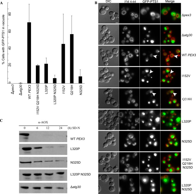
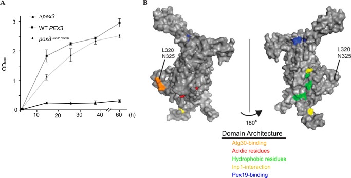
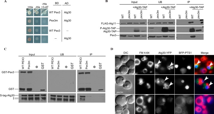
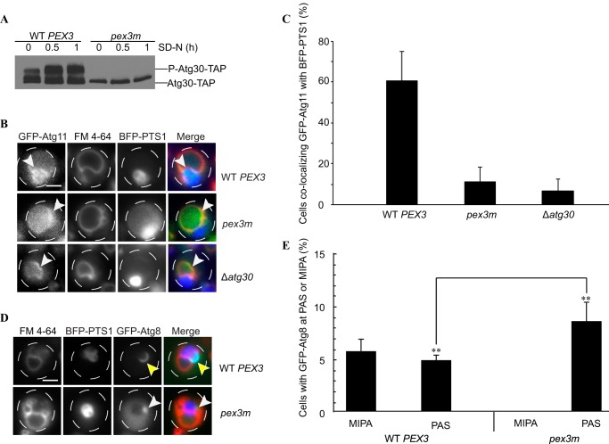
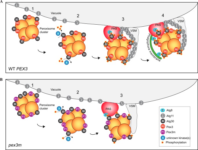
Pexophagy is a process that selectively degrades peroxisomes by autophagy. The Pichia pastoris pexophagy receptor Atg30 is recruited to peroxisomes under peroxisome proliferation conditions. During pexophagy, Atg30 undergoes phosphorylation, a prerequisite for its interactions with the autophagy scaffold protein Atg11 and the ubiquitin-like protein Atg8. Atg30 is subsequently shuttled to the vacuole along with the targeted peroxisome for degradation. Here, we defined the binding site for Atg30 on the peroxisomal membrane protein Pex3 and uncovered a role for Pex3 in the activation of Atg30 via phosphorylation and in the recruitment of Atg11 to the receptor protein complex. Pex3 is classically a docking protein for other proteins that affect peroxisome biogenesis, division, and segregation. We conclude that Pex3 has a role beyond simple docking of Atg30 and that its interaction with Atg30 regulates pexophagy in the yeast P. pastoris.

Keywords: Autophagy; Membrane Trafficking; Peroxisome; Pexophagy; Protein-Protein Interaction; Receptor Regulation; Signaling.


Figures

FIGURE 1.
FIGURE 1.
Pex3 mutants with pexophagy defect. A, the Δpex3 strain expressing a peroxisome reporter (GFP-PTS1) was transformed with plasmids expressing either wild-type PEX3 or mutant pex3 ORFs. Strains Δpex3 and Δatg30 were used as controls. The percentage of cells that contained GFP fluorescence in the vacuole after 3 h in SD−N was estimated (mean % ± S.E. of three experiments). Quantification was done on at least 300 cells of each strain. B, representative images in A showing cells under pexophagy conditions. FM 4-64 was used to stain the vacuolar membrane. Arrowheads indicate GFP fluorescence in the vacuole. Scale bar = 2 μm. DIC, differential interference contrast. C, biochemical pexophagy assay. Cell lysates were prepared as described under “Experimental Procedures” and resolved by SDS-PAGE. Western blotting was performed with antibodies against P. pastoris AOX.
FIGURE 2.
FIGURE 2.
Identification of a novel pexophagy-specific domain. A, strains Δpex3 and Δpex3 complemented with plasmids containing either WT PEX3 or pex3m were evaluated for proper peroxisome proliferation via growth in methanol medium. Mean absorbance ± S.E. from three individual experiments is presented. B, hypothetical predicted structure of P. pastoris Pex3 modeled in PyMOL based on human PEX3. The surface of the protein is visualized in gray, and protein-binding domains are shown in colors as indicated.
FIGURE 3.
FIGURE 3.
Pex3-Atg30 interaction is not required to localize Atg30 to the peroxisome. A, yeast two-hybrid analysis for interaction between Atg30 and WT Pex3 or Pex3m. Atg30 was fused to the Gal4 activation domain (AD), and Pex3 was fused to the Gal4 DNA-binding domain (BD). Cell growth in the absence of histidine was used as a measure of protein-protein interaction. Dashes denote empty plasmids used as controls: pGBKT7 (Gal4 DNA-binding domain) and pGADGH (Gal4 activation domain). B, Δypt7 cells with endogenous PEX3 replaced with WT PEX3-HYGROR or pex3m-HYGROR were transformed with FLAG-Atg11 and Atg30-TAP (denoted as +Atg30-TAP). Immunoprecipitation (IP) was performed using IgG-agarose beads under pexophagy conditions (SD−N). Lysates of cells taken at absorbances of 0.8, 29.2, and 0.8 were loaded as input, immunoprecipitate, and unbound fractions (UB; long exposure), respectively. Immunoprecipitated proteins were visualized with anti-FLAG, anti-calmodulin-binding peptide, and anti-P. pastoris Pex3. P-Atg30 denotes the phosphorylated species. C, His-Atg30 bound to GST-Pex3ΔN, but not to GST-Pex3mΔN or GST, in vitro. There was equivalent loading in the input, unbound fraction, and immunoprecipitate (pulldown) lanes. Ø contained His-Atg30, but no GST species. Immunoblotting was performed with anti-S·Tag (to detect Atg30) and anti-GST antibodies. D, Δpex3 cells were transformed with Atg30-YFP, BFP-PTS1, and plasmids containing either WT PEX3 (upper panels) or pex3m (middle panels) and subjected to peroxisome proliferation and subsequent pexophagy conditions (SD−N, 1 h), followed by the localization of Atg30 to peroxisomal membranes (white arrows). FM 4-64 was used to label the vacuolar membrane. Δpex3 cells did not form peroxisomes, and Atg30 was cytosolic (lower panels). Scale bar = 2 μm.
FIGURE 4.
FIGURE 4.
pex3m affects Atg30 phosphorylation, Atg11 localization, and phagophore formation. A, Δypt7 (WT PEX3-HYGROR or pex3m-HYGROR) cells containing Atg30-TAP were followed after peroxisome induction to monitor the phosphorylation status of Atg30 upon switching the medium to SD−N. P-Atg30-TAP denotes the phosphorylated Atg30 species. B, WT PEX3-HYGROR, pex3m-HYGROR, and Δatg30 cells containing GFP-Atg11, BFP-PTS1, and FM 4-64 were visualized after switching to SD−N (1 h). White arrowheads indicate GFP-Atg11 localization. Scale bar = 2 μm. C, cells from B were quantified for GFP-Atg11 accumulation in the interface of the vacuole (FM 4-64) and peroxisomes (BFP-PTS1) (mean % of three experiments ± S.E.). D, Δpex3 cells were transformed with plasmids containing either WT PEX3 or pex3m along with GFP-Atg8 and BFP-PTS1 to monitor phagophore (MIPA) formation (yellow arrowheads) or PAS organization (white arrowheads) upon induction of pexophagy. Scale bar = 2 μm. E, MIPA and PAS structures from D were quantified (mean % of three experiments ± S.E.). **, p < 0.001.
FIGURE 5.
FIGURE 5.
Model for Atg30 activation by Pex3. A, in WT cells, Atg30 and Pex3 localize at the peroxisomal membrane (step 1). After pexophagy induction, Atg30 is phosphorylated by an unknown kinase(s) in a Pex3-Atg30 interaction-dependent manner (step 2). Atg8 and Atg11 interact with phosphorylated Atg30, leading to PAS formation and Atg11 accumulation at the interface between peroxisomes and vacuolar membranes (step 3). The phagophore (decorated by Atg8) extends from the PAS and engulfs the peroxisome cluster (step 4) to allow for fusion with the vacuolar membranes and peroxisome degradation inside the vacuole. B, in pex3m cells, Atg30 is targeted to the peroxisome in a Pex3-independent manner (step 1). Atg30 is not properly phosphorylated (step 2). Despite being hypophosphorylated, Atg8 is still recruited to the PAS, yet Atg11 does not accumulate in the proximity of the peroxisome cluster (step 3).

References

    1. Tan C. C., Yu J. T., Tan M. S., Jiang T., Zhu X. C., Tan L. (2014) Autophagy in aging and neurodegenerative diseases: implications for pathogenesis and therapy. Neurobiol. Aging 35, 941–957 - PubMed
    1. Feng Y., He D., Yao Z., Klionsky D. J. (2014) The machinery of macroautophagy. Cell Res. 24, 24–41 - PMC - PubMed
    1. Nakatogawa H., Suzuki K., Kamada Y., Ohsumi Y. (2009) Dynamics and diversity in autophagy mechanisms: lessons from yeast. Nat. Rev. Mol. Cell Biol. 10, 458–467 - PubMed
    1. Yang Z., Klionsky D. J. (2009) An overview of the molecular mechanism of autophagy. Curr. Top. Microbiol. Immunol. 335, 1–32 - PMC - PubMed
    1. Till A., Lakhani R., Burnett S. F., Subramani S. (2012) Pexophagy: the selective degradation of peroxisomes. Int. J. Cell Biol. 2012, 512721. - PMC - PubMed

Publication types

LinkOut - more resources