. 2015 Apr 10;290(15):9614-25.
doi: 10.1074/jbc.M114.614628. Epub 2015 Feb 18.

The transcription factor Mesp1 interacts with cAMP-responsive element binding protein 1 (Creb1) and coactivates Ets variant 2 (Etv2) gene expression

Affiliations

The transcription factor Mesp1 interacts with cAMP-responsive element binding protein 1 (Creb1) and coactivates Ets variant 2 (Etv2) gene expression

Xiaozhong Shi et al. J Biol Chem. .

Abstract

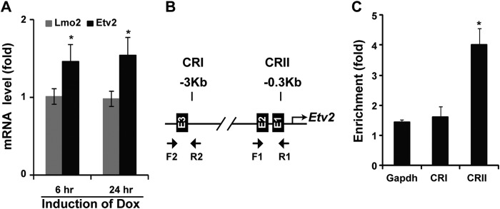
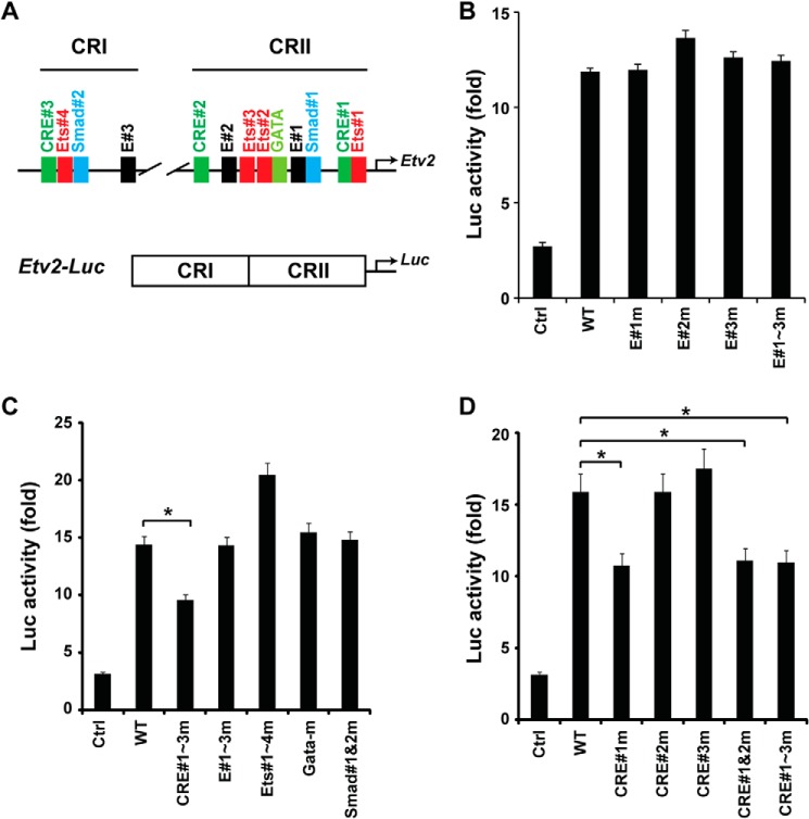
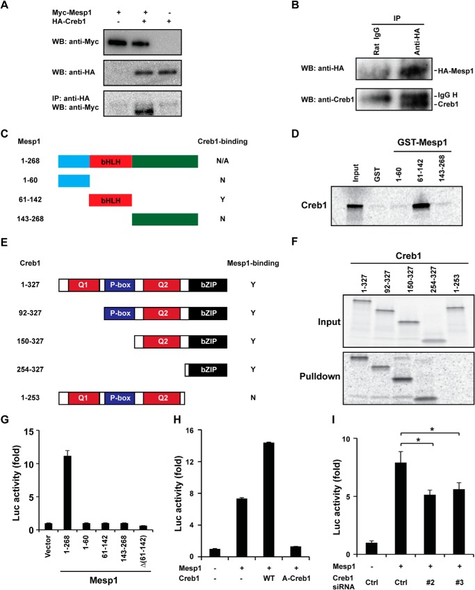
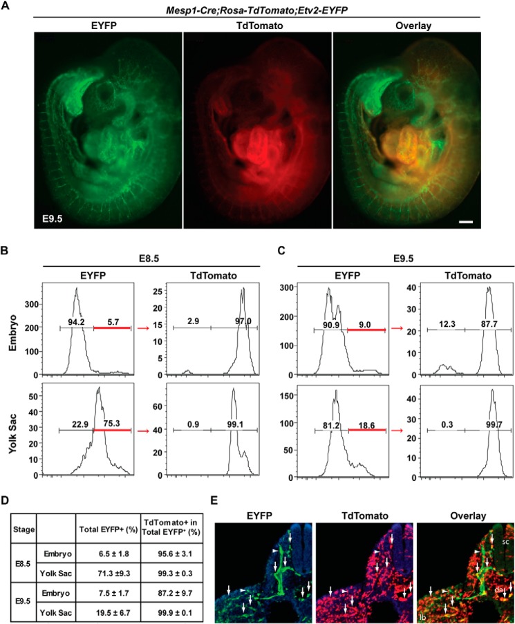
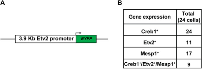
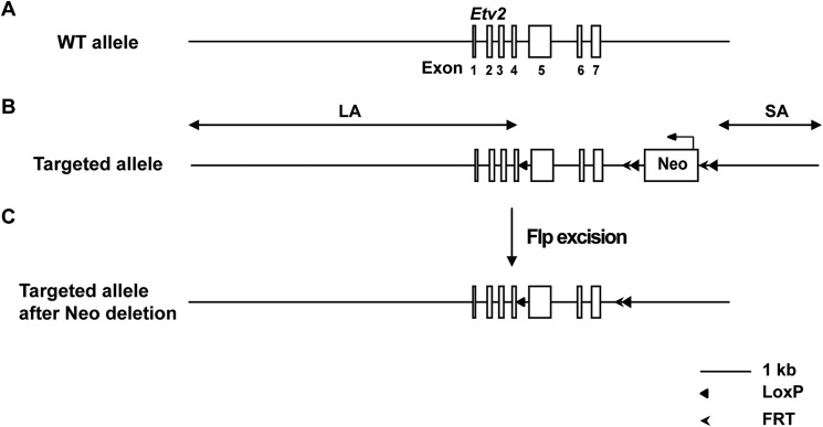
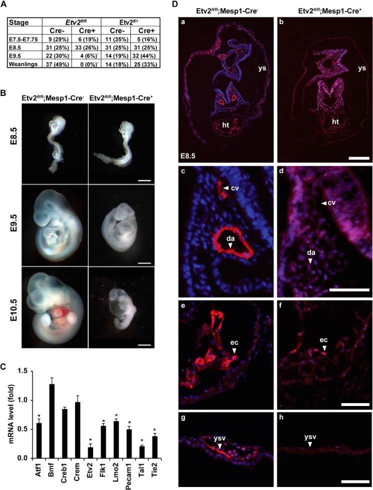
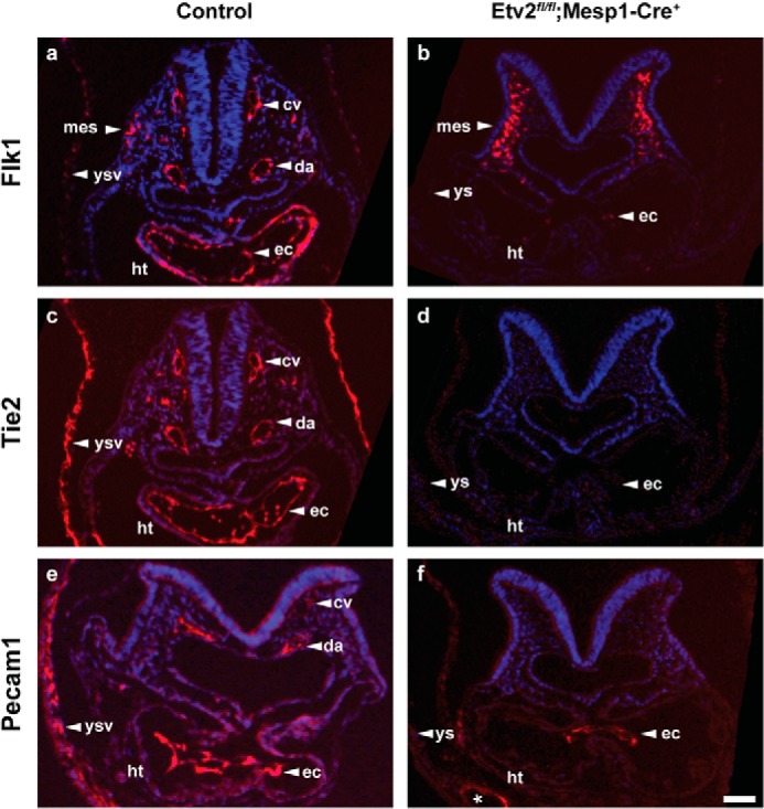
Mesoderm posterior 1 (Mesp1) is well recognized for its role in cardiac development, although it is expressed broadly in mesodermal lineages. We have previously demonstrated important roles for Mesp1 and Ets variant 2 (Etv2) during lineage specification, but their relationship has not been defined. This study reveals that Mesp1 binds to the proximal promoter and transactivates Etv2 gene expression via the CRE motif. We also demonstrate the protein-protein interaction between Mesp1 and cAMP-responsive element binding protein 1 (Creb1) in vitro and in vivo. Utilizing transgenesis, lineage tracing, flow cytometry, and immunostaining technologies, we define the lineage relationship between Mesp1- and Etv2-expressing cell populations. We observe that the majority of Etv2-EYFP(+) cells are derived from Mesp1-Cre(+) cells in both the embryo and yolk sac. Furthermore, we observe that the conditional deletion of Etv2, using a Mesp1-Cre transgenic strategy, results in vascular and hematopoietic defects similar to those observed in the global deletion of Etv2 and that it has embryonic lethality by embryonic day 9.5. In summary, our study supports the hypothesis that Mesp1 is a direct upstream transactivator of Etv2 during embryogenesis and that Creb1 is an important cofactor of Mesp1 in the transcriptional regulation of Etv2 gene expression.

Keywords: Basic Helix-Loop-Helix Transcription Factor (bHLH); ETS Transcription Factor Family; Protein-Protein Interaction; Transcription Regulation; Transcriptional Coactivator.


Figures

FIGURE 1.
FIGURE 1.
Etv2 is a direct downstream target gene of Mesp1. A, Etv2 is induced by Mesp1 using the iMesp1 ES/EB system treated with doxycycline (Dox) for 6 h. The induction is persistent following 24-h treatment. In contrast, Lmo2 is not induced by Mesp1 following 6- or 24-h treatments (*, p < 0.05). B, bioinformatics analysis reveals three E-box motifs in CRI and CRII of the Etv2 upstream 3.9-kb promoter. Specific primers for the ChIP assay are indicated for CRI and CRII. C, Mesp1 binds to the CRI region but not to the CRII region, as revealed using ChIP assays. Gapdh was a negative control for the ChIP assay (*, p < 0.05).
FIGURE 2.
FIGURE 2.
Mesp1 transactivates Etv2 gene expression via the CRE motif. A, top panel, the conserved motifs present in the CRI and CRII regions of the Etv2 3.9-kb promoter. ETS, Ets factor-binding motif; Smad, Smad-binding motif; E, E-box motif; GATA, Gata factor-binding motif. Bottom panel, the Etv2-luc reporter is constructed with CRI and CRII in front of the luciferase gene. B, using transcriptional assays, Mesp1 can transactivate the Etv2-luc reporter up to 12-fold. However, mutation of the E-box motifs, individually or in combination, does not attenuate the transactivation. Ctrl, control. C, CRE motifs are required for Mesp1 transactivation of the Etv2 promoter using mutagenesis screening. Note that mutagenesis of the Ets, Gata, or Smad motifs does not reduce the transactivation by Mesp1 (*, p < 0.05). D, mutation of CRE#1, instead of CRE#2 or CRE#3, attenuates the transactivation by Mesp1. Additional mutations of CRE#2 or CRE#3 motifs do not further enhance the effect of CRE#1 on the transcriptional activation (*, p < 0.05).
FIGURE 3.
FIGURE 3.
Mesp1 interacts directly with Creb1. A, Myc-Mesp1 and HA-Creb1 are overexpressed in C2C12 cells. Overexpression of Myc-Mesp1 and HA-Creb1 is detected using Western blot analysis and anti-Myc or anti-HA sera, respectively. Myc-Mesp1 is coimmunoprecipitated (IP) with HA-Creb1 using a HA antibody (Western blot (WB), anti-Myc). B, HA-Mesp1 is overexpressed to a similar level of endogenous Mesp1 in EBs on day 4. HA-Mesp1 is immunoprecipitated successfully by anti-HA (top panel). Endogenous Creb1 is detected using an anti-Creb serum (bottom panel). C, the deletional constructs of Mesp1. N, no; Y, yes; N/A, not available. D, 35S-labeled Creb1 is pulled down by GST-Mesp1 (61–142) but not Mesp1 (1–60) or Mesp1 (143–268), as summarized in C. E, the Creb1 deletional construct. Q1 and Q2, Glu-rich domains 1 and 2; P-box, phosphorylation domain. F, deletional Creb1 constructs were translated in vitro in the presence of [35S]methione (Input) and then utilized for a pulldown assay with GST-Mesp1 (61–142). All of the deletions harboring the bZIP domain can be pulled down, whereas the constructs lacking the bZIP domain cannot be pulled down, as summarized in E. G, transcriptional activity of Mesp1 deletions. Full-length Mesp1 transactivates the Etv2 reporter, whereas each domain of Mesp1 or deletion of bHLH domain Δ(61–142) does not transactivate the Etv2 reporter. H, wild-type Creb1 augments the activity of Mesp1. The dominant negative inhibitor A-Creb1 represses the activity of Mesp1 to the baseline level. WT, wild-type Creb1. I, knockdown of Creb1 by siRNA #2 and #3 reduces the activity of Mesp1. Ctrl, control, referring to the RNA-induced silencing complex-free siRNA (*, p < 0.05).
FIGURE 4.
FIGURE 4.
The majority of Etv2-EYFP-expressing cells at E8.5-E9.5 arise from Mesp1-Cre+ cells. A, whole-mount images of Mesp1-Cre; Rosa-TdTomato; Etv2-EYFP compound embryos at E9.5. Scale bar = 200 μm. B, FACS analysis of lineage-traced Mesp1-Cre+ cells and Etv2-EYFP+ cells at E8.5 and (C) E9.5 in both the yolk sac and embryo. D, total EYFP+ cells (percent) and TdTomato+ in the total Etv2-EYFP+ population (percent) at E8.5 and E9.5. Note that 95.6% of Etv2-EYFP+ cells at E8.5 and 87.2% of Etv2-EYFP+ cells at E9.5 are derived from cells that have expressed Mesp1-Cre. Note that, in the yolk sac, 98% of Etv2-EYFP+ cells at E8.5 and E9.5 are descendants from Mesp1-Cre+ cells (n = 4). E, immunostaining of EYFP (green, left panel) and TdTomato (red, center panel) in the Mesp1-Cre;Rosa-TdTomato;Etv2-EYFP embryo. The coexpression of EYFP and TdTomato is shown as the overlay (yellow, right panel). The arrows indicate double-positive cells, and the arrowheads point to GFP single positives. sc, spinal cord; da, dorsal aorta; lb, limb bud. The section is at the forelimb level.
FIGURE 5.
FIGURE 5.
Coexpression of Etv2 and Mesp1. A, the 3.9-kb Etv2 promoter-EYFP construct utilized in the A2lox ES construction. B, gene expression of Creb1, Etv2, and Mesp1 in the 24 cells sorted from the reporter cell line at EB day 4. Creb1 was detected in all 24 cells, Etv2 in 11 cells, and Mesp1 in 17 cells. These three genes were detected in 9 of 24 cells (37.5%).
FIGURE 6.
FIGURE 6.
Conditional knock-out of Etv2. A, the seven exons of the Etv2 gene are schematized. B, the conditional targeted allele of Etv2. One LoxP site was placed between exons 4 and 5. The neomycin selection cassette (Neo) is flanked with the FRT-LoxP sequences and placed downstream of exon 7. The schematic of Etv2 conditional knockout reveals a 6.4-kb long arm of homology (LA), a 74-bp LoxP cassette, a 1.85-kb target region (including exons 5–7 and a neomycin cassette flanked with FRT and LoxP), and a 2.02-kb short arm of homology (SA).
FIGURE 7.
FIGURE 7.
Etv2fl/fl;Mesp1-Cre+ embryos are nonviable, and most of them die by E9.5 because of vascular and hematopoietic defects. A, genotypes at E7.5, E8.5, and E9.5 and weanlings from the respective breeding. Note the significant reduction of the Etv2fl/fl; Mesp1-Cre+ embryos at E9.5. *, p < 0.001. B, whole-mount analysis of E8.5-E10.5 embryos. Note the reduced size of the Etv2fl/fl;Mesp1-Cre+ embryos and the pericardial edema and anemia at E10.5 compared with the littermate controls. Scale bars = 500 μm. C, gene expressions in the Etv2fl/fl; Mesp1-Cre+ mutant embryos at E8.5. Note that both endothelial and hematopoietic genes are down-regulated, whereas the expression of Creb1 and Crem is unaffected. Bmf serves as a control. *, p < 0.05. D, histological analysis of Etv2fl/fl; Mesp1-Cre and Etv2fl/fl; Mesp1-Cretg/+ Etv2fl/fl; Mesp1-Cre+ mouse embryos at E8.5 stained with the endomucin antibody (a–h) reveals the presence of endothelial lineages in control embryos that are largely absent in the Etv2fl/fl; Mesp1-Cre+ mutant embryos. Shown are representative sections of dorsal aortae and cardinal veins (c and d), endocardium (e and f), and yolk sac vasculature (g and h). cv, cardinal vein; da, dorsal aorta; ec, endocardium; ht, heart; ys, yolk sac; ysv, yolk sac vasculature. Scale bars = 200 μm (a and b) and 50 μm (c–h).
FIGURE 8.
FIGURE 8.
Control embryos (a and c, Etv2fl/+; Mesp1-Cre; e, Etv2fl/fl; Mesp1-Cre) and an Etv2fl/fl;Mesp1-Cre+ conditional mutant embryo (b, d, and f) at E8.5 were sectioned transversely at the heart level and immunostained for antibodies against Flk1 (a and b), Tie2 (c and d), and Pecam1 (e and f). The asterisk (f) indicates occasional vessel-like structures in the yolk sac that are Pecam1-positive. Note that the majority of endothelial lineages are missing in the Etv2fl/fl;Mesp1-Cre+ conditional mutants. cv, cardinal vein; da, dorsal aorta; ec, endocardium; ht, heart; mes, mesenchyme, ys, yolk sac; ysv, yolk sac vasculature. Scale bar = 50 μm.

References

    1. Keller G., Lacaud G., Robertson S. (1999) Development of the hematopoietic system in the mouse. Exp. Hematol. 27, 777–787 - PubMed
    1. Ferkowicz M. J., Yoder M. C. (2005) Blood island formation: longstanding observations and modern interpretations. Exp. Hematol. 33, 1041–1047 - PubMed
    1. Li W., Ferkowicz M. J., Johnson S. A., Shelley W. C., Yoder M. C. (2005) Endothelial cells in the early murine yolk sac give rise to CD41-expressing hematopoietic cells. Stem. Cells Dev. 14, 44–54 - PubMed
    1. Wilson N. K., Foster S. D., Wang X., Knezevic K., Schütte J., Kaimakis P., Chilarska P. M., Kinston S., Ouwehand W. H., Dzierzak E., Pimanda J. E., de Bruijn M. F., Göttgens B. (2010) Combinatorial transcriptional control in blood stem/progenitor cells: genome-wide analysis of ten major transcriptional regulators. Cell Stem Cell 7, 532–544 - PubMed
    1. Moignard V., Woodhouse S., Fisher J., Göttgens B. (2013) Transcriptional hierarchies regulating early blood cell development. Blood Cells Mol. Dis. 51, 239–247 - PubMed

Publication types

MeSH terms

Substances