Review
. 2015 Jul;26(7):1503-12.
doi: 10.1681/ASN.2014101000. Epub 2015 Feb 18.

Current Understanding of the Role of Complement in IgA Nephropathy

Affiliations
Review

Current Understanding of the Role of Complement in IgA Nephropathy

Nicolas Maillard et al. J Am Soc Nephrol. 2015 Jul.

Abstract

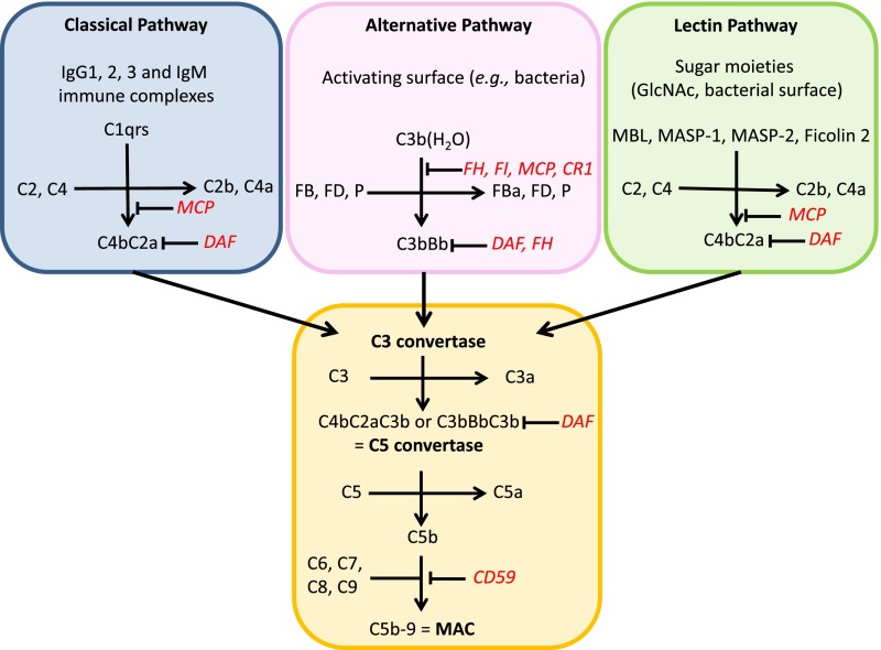
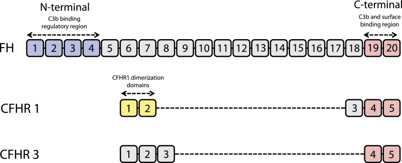
Complement activation has a role in the pathogenesis of IgA nephropathy, an autoimmune disease mediated by pathogenic immune complexes consisting of galactose-deficient IgA1 bound by antiglycan antibodies. Of three complement-activation pathways, the alternative and lectin pathways are involved in IgA nephropathy. IgA1 can activate both pathways in vitro, and pathway components are present in the mesangial immunodeposits, including properdin and factor H in the alternative pathway and mannan-binding lectin, mannan-binding lectin-associated serine proteases 1 and 2, and C4d in the lectin pathway. Genome-wide association studies identified deletion of complement factor H-related genes 1 and 3 as protective against the disease. Because the corresponding gene products compete with factor H in the regulation of the alternative pathway, it has been hypothesized that the absence of these genes could lead to more potent inhibition of complement by factor H. Complement activation can take place directly on IgA1-containing immune complexes in circulation and/or after their deposition in the mesangium. Notably, complement factors and their fragments may serve as biomarkers of IgA nephropathy in serum, urine, or renal tissue. A better understanding of the role of complement in IgA nephropathy may provide potential targets and rationale for development of complement-targeting therapy of the disease.

Keywords: IgA nephropathy; complement; immune complexes.


Figures

Figure 1.
Figure 1.
Three pathways of complement activation. The classical pathway is triggered by IgG– and/or IgM–containing immune complexes. The alternative pathway is constantly initiated by spontaneous hydrolysis of C3 [C3b(H2O)] that is efficiently powered by the covalent attachment of C3b on an activating surface. The lectin pathway requires a particular sugar moiety pattern (N-acetylglucosamine [GlcNAc]) to be recognized and bound by MBL, leading to a classical pathway–like activation cascade. Each pathway leads to formation of a C3 convertase. The addition of C3b to the C3 convertase creates a C5 convertase that, in turn, triggers the assembly of the membrane attack complex (C5b-9), which is also known as the terminal pathway complete complex. Regulatory factors are in red. CR1, complement receptor 1; FD, factor D; MAC, membrane attack complex; MCP, membrane cofactor protein; P, properdin.
Figure 2.
Figure 2.
C3 proteolytic cascade. The hydrolysis of C3 leads to the release of activation products C3a, a potent anaphylatoxin, and C3b, which contains a highly reactive thioester bond that can covalently bind activating surfaces, such as a bacterial wall. This attachment of C3b initiates a powerful amplification system from the formation of the C3 convertase to the subsequent proteolysis of new molecules of C3, allowing more C3b to bind to the surface. This amplification is controlled by regulator molecules, such as FI, FH, and complement receptor 1 (CR1), that degrade C3b into products that cannot contribute to the formation of the C5 convertase. Detection of these inactive breakdown products (iC3b, C3c, C3dg, and C3d) is considered evidence of activation of C3. The numbers, in kilodaltons, represent the molecular masses of the corresponding polypeptides. MCP, membrane cofactor protein.
Figure 3.
Figure 3.
Comparison of the structures of FH, CFHR1, and CFHR3. The structure of FH contains 20 SCRs. SCR1–SCR4 possess the regulatory activity (FI cofactor activity and decay acceleration of the C3 convertase) as well as a weak C3b-binding capacity. SCR19 and SCR20 contain the most powerful C3b-binding zone that is critical for binding to cell surfaces. This latter area is the hotspot of aHUS-associated mutations, likely explained by the loss of ability of an abnormal FH to bind to endothelial cells. CFHR1 and CFHR3 resemble FH by the presence of structurally similar SCRs. These molecules possess the corresponding C3b/cell surface–binding zone (SCR4 and SCR5 corresponding to SCR19 and SCR20, respectively, of FH) but lack the regulatory portion (having no region equivalent to SCR1–SCR4). In contrast to FH and CFHR3, CFHR1 contains a unique pair of highly conserved SCR1 and SCR2 (also shared with CFHR2 and CFHR5). These domains enable formation of homo- and heterodimers involving CFHR1, CFHR2, and CFHR5.
Figure 4.
Figure 4.
Proposed mechanism to explain the protective effect of CFHR1,3 deletion on the development of IgAN. CFHR1 and CFHR3 proteins can bind to C3b in competition with FH. The regulatory activities of CFHR1 and CFHR3 are less efficient than those of FH. CFHR1,3 deletion, thus, allows FH to bind C3b effectively and thereby to strongly inhibit the initiation and amplification of the alternative pathway cascade.
Figure 5.
Figure 5.
Integrative view of the role of complement activation in the four-hit model of the pathogenesis of IgAN. C3 can be activated directly by IgA1–containing immune complexes formed from Gd-IgA1 and antiglycan antibodies and increase the pathogenic potential of these complexes. Other proteins can bind Gd-IgA1, such as the soluble form of Fcα receptor (sCD89), to generate complexes with Gd-IgA1. An association between the levels of sCD89-IgA complexes in serum and the severity of IgAN has been observed. Specifically, patients with IgAN without disease progression had high levels of sCD89 in contrast to low levels of sCD89 in the disease progression group, suggesting that sCD89-IgA complexes may be protective. In contrast, an animal model suggested that interaction between four entities—Gd-IgA1, sCD89, transferrin receptor, and transglutaminase 2 in mesangial cells—is needed for disease development. The lectin and alternative pathways can each contribute to the glomerular damage induced by immune complexes in the mesangium. Mesangial cells can also play an active role, arising from their capacity to be stimulated by C3a as well as produce C3 in response to an inflammatory stimulus.

References

    1. Berger J, Hinglais N: Intercapillary deposits of IgA-IgG. J Urol Nephrol (Paris) 74: 694–695, 1968 - PubMed
    1. Berthoux FC, Mohey H, Afiani A: Natural history of primary IgA nephropathy. Semin Nephrol 28: 4–9, 2008 - PubMed
    1. Conley ME, Cooper MD, Michael AF: Selective deposition of immunoglobulin A1 in immunoglobulin A nephropathy, anaphylactoid purpura nephritis, and systemic lupus erythematosus. J Clin Invest 66: 1432–1436, 1980 - PMC - PubMed
    1. Russell MW, Mestecky J, Julian BA, Galla JH: IgA-associated renal diseases: Antibodies to environmental antigens in sera and deposition of immunoglobulins and antigens in glomeruli. J Clin Immunol 6: 74–86, 1986 - PubMed
    1. Jennette JC: The immunohistology of IgA nephropathy. Am J Kidney Dis 12: 348–352, 1988 - PubMed

Publication types

Substances

LinkOut - more resources