. 2015 Mar 11;43(5):2543-59.
doi: 10.1093/nar/gkv083. Epub 2015 Feb 18.

bHLH proteins involved in Drosophila neurogenesis are mutually regulated at the level of stability

Affiliations

bHLH proteins involved in Drosophila neurogenesis are mutually regulated at the level of stability

Marianthi Kiparaki et al. Nucleic Acids Res. .

Abstract

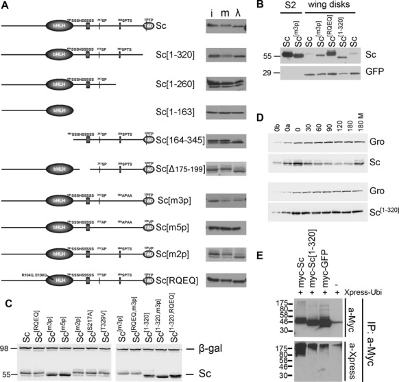
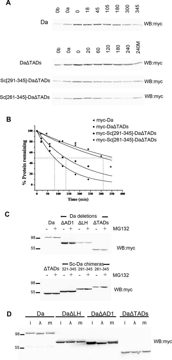
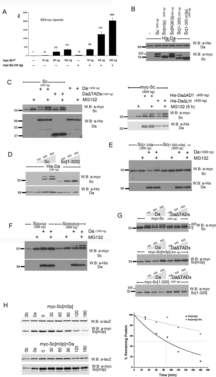
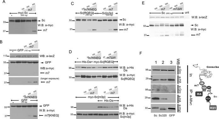
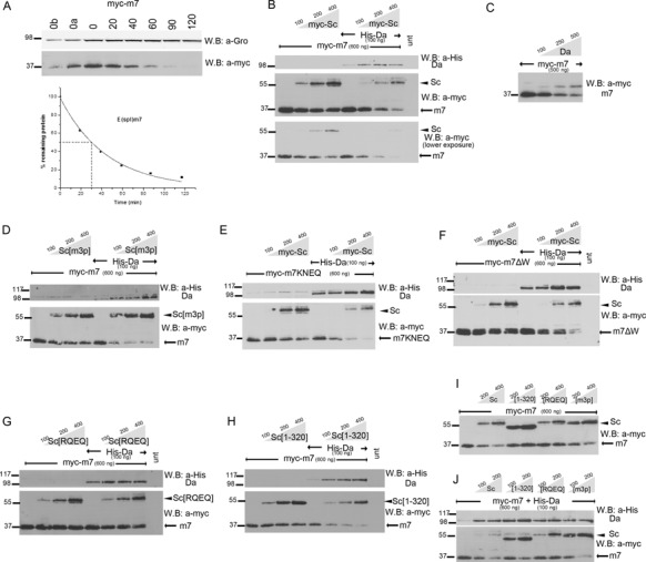
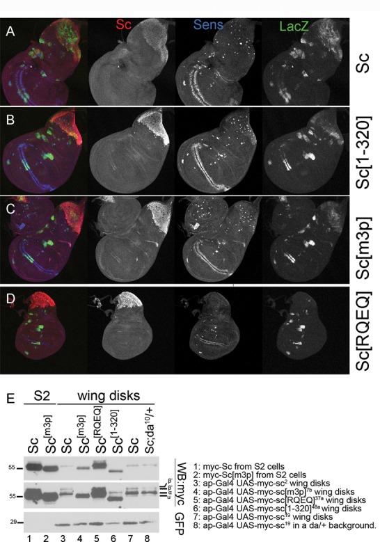
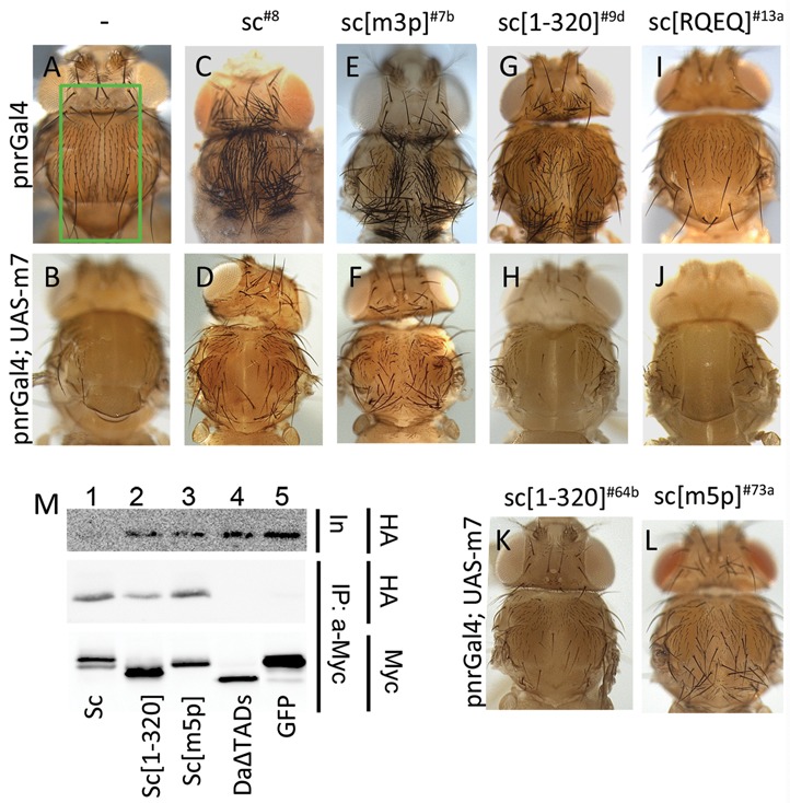
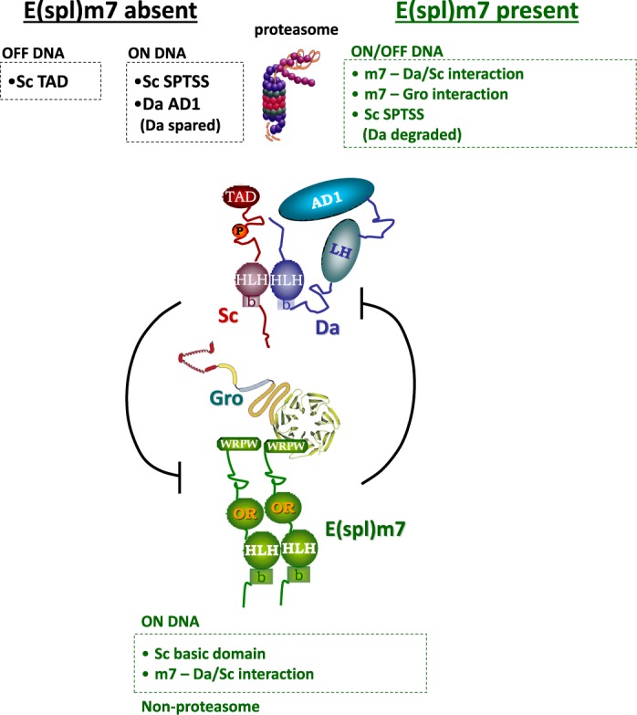
Proneural bHLH activators are expressed in all neuroectodermal regions prefiguring events of central and peripheral neurogenesis. Drosophila Sc is a prototypical proneural activator that heterodimerizes with the E-protein Daughterless (Da) and is antagonized by, among others, the E(spl) repressors. We determined parameters that regulate Sc stability in Drosophila S2 cells. We found that Sc is a very labile phosphoprotein and its turnover takes place via at least three proteasome-dependent mechanisms. (i) When Sc is in excess of Da, its degradation is promoted via its transactivation domain (TAD). (ii) In a DNA-bound Da/Sc heterodimer, Sc degradation is promoted via an SPTSS phosphorylation motif and the AD1 TAD of Da; Da is spared in the process. (iii) When E(spl)m7 is expressed, it complexes with Sc or Da/Sc and promotes their degradation in a manner that requires the corepressor Groucho and the Sc SPTSS motif. Da/Sc reciprocally promotes E(spl)m7 degradation. Since E(spl)m7 is a direct target of Notch, the mutual destabilization of Sc and E(spl) may contribute in part to the highly conserved anti-neural activity of Notch. Sc variants lacking the SPTSS motif are dramatically stabilized and are hyperactive in transgenic flies. Our results propose a novel mechanism of regulation of neurogenesis, involving the stability of key players in the process.


Figures

Figure 1.
Figure 1.
Phosphorylation and stability of Sc. In all panels N-terminally 3xmyc-tagged Sc proteins were detected by anti-Myc tag antibody. (A) The schematic depicts the Sc protein, highlighting its bHLH domain, a Ser-rich stretch and three (S/T)P conserved motifs in the C-terminal half. A series of deletion and point mutations are shown. Next to each the western blot shows (i) the Sc variant expressed in S2 cells (input), (m) the S2 cell extract mock-treated with λ-phosphatase buffer and (λ) the same extract treated with λ-phosphatase. An increase in migration indicates dephosphorylation. (B) Western blot containing extracts from Sc-transfected S2 cells or Sc expressing wing disks (ap-Gal4; UAS-GFP UAS-myc-sc). Note that Sc[m3p] migrates faster than Sc(wt) both in S2 cells and in fly tissue. GFP is shown as a loading control. (C) A comparison of all Sc variants from S2 cells co-transfected with Ract-myc-sc and Ract-lacZ; the β-galactosidase protein is detected as a loading control. Sc[m3p], Sc[m5p] and the versions truncated after residue 320 (lacking the acidic C-terminal TAD) accumulate to higher levels. Also note the faster migration of the phosphomutants (m3p and m5p). (D) Degradation kinetics of transfected myc-Sc and myc-Sc[1–320] after protein synthesis inhibition by cycloheximide. Endogenous Groucho (a stable protein) is used as a loading control. 0, 30,…, 180 refer to minutes after cycloheximide addition. 180M: 180 min after cycloheximide+MG132 addition. 0a, 0b: 1/2 or 1/4, respectively, of input (0 min) for densitometry calibration. One indicative experiment is shown out of four (Sc) or five (Sc[1–320]) repeats. (E) The indicated myc-tagged protein expression constructs were co-transfected with Xpress-tagged ubiquitin. Ubiquitylated species were detected after IP with anti-Myc and western blot (WB) with anti-Xpress (bottom panel). The top panel shows an anti-Myc WB to estimate the amount of Myc-tagged protein in each IP. Cells had been treated with MG132 (for 5 h) before lysis. Note higher level of ubiquitin signal in the Sc lane versus Sc[1–320] and GFP.
Figure 2.
Figure 2.
Stability and phosphorylation of Da. In all panels N-terminally 3xmyc-tagged Da proteins were detected. (A) Turnover kinetics of Da and three variants after protein synthesis block. Lanes are labeled as in Figure 1D. (B) The densitometry plots from the blots of panel B. Each measurement was repeated 2–3 times; one indicative experiment is shown. The estimated half-lives (number of repeats in parenthesis) are: Da 275±90 min (n = 3), DaΔTADs > 240 min (n = 3), Sc[291–345]-DaΔTADs 141±43 (n = 2), Sc[261–345]-DaΔTADs 83±10 (n = 2). (C) Da proteins detected from transfected S2 cells treated (+) or not (-) with MG132 for 5 h before lysis. Proteasome inhibition does not seem to increase the levels of Da or versions deleted for one or both of its TADs. Instead, it detectably increased the levels of Da–Sc chimeras bearing the Sc TAD and nearby residues. (D) λ-phosphatase treatment of S2 cell extracts expressing the indicated Da variants. i: input, λ: treated, m: mock-treated. Note the increase in migration for myc-Da (wt) and myc-DaΔAD1, but not myc-DaΔLH or myc-DaΔTADs.
Figure 3.
Figure 3.
Proteasomal degradation of Sc in the presence of Da depends on phosphorylation of Sc, its DNA binding ability and on the AD1 domain of Da. (A) Luciferase assays on transiently transfected S2 cells with an EE4-luc reporter. Activation levels by increasing amounts of myc-Sc in the presence or absence of co-transfected Da. Relative luciferase units (rlu) are shown and are normalized on the activity of the reporter gene alone, set to 1. Averages/standard deviations of triplicates are shown. In all subsequent panels N-terminally 3xmyc-tagged Sc proteins or His-tagged Da were detected. The amount loaded was adjusted according to the activity of luciferase expressed from control plasmids (Ract-luc) transfected at the same time as the indicated plasmids (to measure transfection efficiency). (B) Whole cell extracts from S2 cells co-transfected with expression constructs as indicated. Note that Da co-expression causes the appearance of new Sc bands that run about 5 kd above the major band. The triple phosphomutant (m3p) abolishes this modification. (C) S2 cells transfected with the indicated constructs were treated (+) or not (-) with MG132 (for ∼5 h) before lysis. Proteasome inhibition increased the levels of Sc when it was expressed alone or together with Da. However, upon co-expression with DaΔTADs, only the β/β’ forms showed significant stabilization. Proteasome inhibition did not seem to increase the levels of Da (lower panel). The same experiment was done using deletion variants DaΔAD1 and DaΔLH (right panel). Proteasome inhibition increased the levels of Sc when it was expressed alone or together with DaΔLH. However, upon co-expression with DaΔAD1, only the β/β’ forms showed significant stabilization. Proteasome inhibition did not seem to increase the levels of Da variants (lower panel). (D) Blots from S2 cell extracts transfected with Da and increasing amounts of Sc or Sc[1–320]. Note that increasing Sc or Sc[1–320] did not diminish the levels of Da; if anything, it caused a small upregulation. (E,F) Cells transfected with Sc[1–320], Sc[1–320, m3p], Sc[m3p] and Sc[RQEQ] in the absence or presence of His-Da were treated or not with MG132. Note that proteasome inhibition stabilized solo Sc[m3p] and Sc[RQEQ], but had no effect when Da is co-expressed (D). Sc[1–320] solo was barely stabilized by MG132 treatment, but when Da was co-expressed MG132 stabilized Sc[1–320]. Finally, MG132 did not seem to increase the levels of Sc[1–320, m3p], whether expressed alone or with Da. (G) Steady-state levels of Sc, Sc[m3p] or Sc[1–320] with increasing amounts of Da variants. Sc[m3p] levels increased with increasing Da or DaΔTADs. Sc[1–320] levels dropped with increasing Da, but were not affected by DaΔTADs. For comparison, Sc levels were not significantly affected by either Da or DaΔTADs; if anything, a weak stabilization at low Da levels was observed. Note that the β form is much more prominent for Sc than for Sc[1–320] and is absent in Sc[m3p]. (H) Degradation kinetics of myc-Sc[m3p] transfected alone or co-transfected with Da after protein synthesis inhibition by cycloheximide. Co-transfected β-galactosidase (lacZ, a stable protein) was used as a loading control. 0, 30,…, 180 refer to minutes after cycloheximide addition. 0a, 0b: 1/2 or 1/4, respectively, of input (0 min) for densitometry calibration. One indicative blot and densitometry plot is shown for each condition. The estimated half-lives are (number of repeats in parenthesis): Sc[m3p] 75±4 min (n = 4), Sc[m3p]/Da 230±130 min (n = 2).
Figure 4.
Figure 4.
Scute and Da degradation by E(spl)m7 is independent of DNA binding, but depends on m7–Gro interaction and the Sc SPTSS motif. (A–E) Whole cell extracts from S2 cells co-transfected with expression constructs as indicated. In all panels N-terminally 3xmyc-tagged Sc or E(spl)m7 proteins or His-tagged Da were detected. The amount loaded was adjusted according to the activity of luciferase (or β-galactosidase, for blot 4E) expressed from control plasmids (Ract-luc or Ract-lacZ) transfected at the same time as the indicated plasmids (to measure transfection efficiency). In the anti-myc blots, the upper band (arrowhead) is myc-Sc or myc-GFP and the lower (arrow) is myc-m7. (A) Increasing levels of m7 led to degradation of Sc. (B) The same effect was not observed for GFP. (C) Increasing levels of m7KNEQ degraded Sc[RQEQ]; whereas increasing levels of m7 did not degrade Sc[m3p]. (D) Increasing levels of m7KNEQ could degrade Da together with Sc[RQEQ]; whereas m7ΔW had impaired ability to do so. Also Sc[m3p] could not be degraded by m7, even in the presence of Da, which was diminished by increasing levels of m7 (lower panel). Asterisk in A and D: half amount of the adjacent sample is loaded for quantity validation. One hundred nanogram of Gro expressing plasmid was co-transfected in experiments 4E and 4D. (E) myc-tagged proteins (Sc, Sc[1–320] and GFP, as indicated) were immunoprecipitated from S2 cells. Immunoprecipitation efficiency (a-Myc panel) and the presence of coprecipitated HA-tagged m7 (a-HA panel) and endogenous Gro (a-Gro) were assayed. Input for the levels of endogenous Gro and the co-transfected HA-m7 are shown in the two upper panels (In). HA-m7 interacts strongly with myc-Sc and weakly with myc-Sc[1–320]. We had shown earlier that, although the major interaction domain for E(spl)m7 is the Sc C-terminal TAD, a weaker interaction exists with the Sc[1–260] fragment (45). In the case of the strong complex formation (Sc–m7), Gro is also co-immunoprecipitated, showing the existence of a super-complex as it shown in the schematic.
Figure 5.
Figure 5.
Degradation of E(spl)m7 is enhanced by Scute and Da proteins. (A) Degradation kinetics of transfected myc-m7 after protein synthesis inhibition by cycloheximide. 0, 20,…, 120 refer to minutes after cycloheximide addition. 0a, 0b: 1/2 or 1/4, respectively, of input (0 min) for densitometry calibration. Myc-tagged protein levels were estimated by densitometry and normalized against endogenous Gro. Graph shows the estimated protein levels at each time-point and the half-life of myc-m7. The experiment was repeated three times; only one is shown for clarity. The estimated half-life was 26±2 min. B-J: Whole cell extracts from S2 cells transfected with expression constructs as indicated. The amount loaded was adjusted according to the activity of luciferase (or β-galactosidase, for blot 5C) expressed from control plasmids transfected at the same time as the indicated plasmids (to control for variability in transfection efficiency). In the anti-myc blots, the upper band (arrowhead) is myc-Sc and the lower (arrow) is myc-m7. (B) Increasing Sc expression reduces m7 levels, especially upon Da co-expression. (C) Da alone can reduce m7 levels. Note that it also causes the accumulation of a high MW form of m7. Although m7 is monoubiquitylated (see Supplementary Figure S6A), the monoubiquitylated form did not increase upon Da co-expression (data not shown). (D) The Sc SPTSS motif is dispensable for m7 degradation. (E) m7 DNA binding or (F) Gro binding are dispensable for its degradation. (G) Sc DNA binding is needed for m7 degradation. (H) The Sc TAD is dispensable for m7 degradation. (I,J) Side-by-side comparison of m7 degradation promoted by different Sc variants. Sc[RQEQ] is the least active. Sc[1–320] is also less active than wt Sc, especially in the absence of co-expressed Da; this may be due to inability to get recruited onto m7.
Figure 6.
Figure 6.
Differential activity and stability of Sc variants in vivo. (A-D) All panels show wing disks carrying one copy of the EE4-lacZ reporter and overexpressing the indicated myc-tagged Sc variants in the pnr domain (pnrGal4 driver). Immunostaining: Sc (anti-myc, red), β-galactosidase (anti-β-gal, green) and Sens, a SOP marker, (anti-Sens, blue). Note that Sc[m3p] and Sc[1–320] produced a higher number of SOPs than wt Sc. The transgenic lines used for Sc[1–320] (line 48a) and Sc[m3p] (line 7b) cause pupal/pharate lethality, whereas expression of UAS-myc-sc (line 2) or UAS-myc-sc[RQEQ] (line 13a) do not affect viability. (E) (Same blot as Figure 1B with a longer exposure added.) Western blot containing extracts from Sc-transfected S2 cells or Sc expressing wing disks (ap-Gal4; UAS-GFP UAS-myc-sc), as indicated. The transgenic lines used are listed next to the blot. UAS-GFP was co-expressed in all wing disk samples and is detected for quantitative comparison. Sc[RQEQ], Sc[1–320] and Sc[m3p] accumulated to higher levels than Sc (wt). Note that the Da-dependent modifications of Sc (the β/β’ bands) were very pronounced in wing disks, suggesting that Da is expressed in relatively high endogenous levels in this tissue. Upon halving the dose of da+ (last lane), we noticed that the α band was enhanced while β/β’ were reduced.
Figure 7.
Figure 7.
Phosphorylation of Sc in vivo modulates its susceptibility to E(spl) m7 repression. (A, C, E, G, I) Thoraces from pnr-Gal4 flies overexpressing the indicated Sc variants. The pnr expression domain is boxed in panel A. Note the production of ectopic bristles by all Sc variants, except Sc[RQEQ], where mild bristle loss is seen (I). C (UAS-myc-sc8) and E (UAS-myc-sc[m3p]7b) are pharate escapers. G (UAS-myc-sc[1–320]9d) is the only viable line with pnr-Gal4 and I (UAS-myc-sc[RQEQ]13a) is viable, as are all Sc[RQEQ] lines. (B, D, F, H, J) Thoraces from pnr-Gal4 flies overexpressing the indicated Sc variants along with E(spl)m7. m7 strongly inhibits ectopic bristle production, with the exception of panel F, where a large number of bristles persist. (K) pnr-Gal4; UAS-m7. UAS-myc-sc[1–320]64b; line 64b never gives pharate escapers when expressed alone (without m7) and produces many more SOPs in larvae than line 9d (G). Still it is more effectively suppressed by UAS-m7 than Sc[m3p] (F). (L) pnr-Gal4; UAS-m7. UAS-myc-sc[m5p]73a; line 73a never gives pharate escapers when expressed alone (without m7) but is less susceptible than the equally inviable Sc[1–320]64b to m7 overexpression. (M) myc-tagged proteins (as indicated) were expressed and immunoprecipitated from S2 cells. Immunoprecipitation efficiency (Myc, lower panel) and the presence of coprecipitated HA-tagged m7 (HA, middle panel) were assayed. Upper panel shows the levels of the HA-m7 in the cell extracts (input). All Sc variants interact with HA-m7. GFP, the negative control and DaΔTADs (lacks the AD1 and LH domains) do not immunoprecipitate HA-m7.
Figure 8.
Figure 8.
Summary of the main features of Sc and E(spl)m7 degradation. In the absence of E(spl)m7 (black font) Sc can be degraded by two proteasome-dependent mechanisms, one that predominates in the presence of Da (the on-DNA mechanism) and the other that predominates in the absence of Da (the off-DNA mechanism). In the off-DNA mode the most important degron is the C-terminal TAD of Sc. In the on-DNA mechanism, the SPTSS motif becomes more important. Also of great importance is the Da AD1 motif, which acts as a trans-degron for Sc, as Da itself is spared from degradation. In the presence of E(spl)m7 (green font), degradation can take place either on or off DNA and requires the recruitment of Gro onto the Sc–(Da)–m7 complex. The SPTSS motif is also important. In this instance, Da is also degraded. Reciprocally Da/Sc stimulate the turnover of E(spl)m7, which happens on DNA, as it needs the Sc basic domain. Besides the Sc basic domain, only the ability of Da/Sc to recruit E(spl)m7 is needed for m7 degradation, which takes place via a non-proteasomal pathway.

Similar articles

Cited by

References

    1. Massari M.E., Murre C. Helix-loop-helix proteins: regulators of transcription in eucaryotic organisms. Mol. Cell. Biol. 2000;20:429–440. - PMC - PubMed
    1. Villares R., Cabrera C.V. The achaete-scute gene complex of D. melanogaster: conserved domains in a subset of genes required for neurogenesis and their homology to myc. Cell. 1987;50:415–424. - PubMed
    1. Guillemot F., Lo L.C., Johnson J.E., Auerbach A., Anderson D.J., Joyner A.L. Mammalian achaete-scute homolog 1 is required for the early development of olfactory and autonomic neurons. Cell. 1993;75:463–476. - PubMed
    1. Bertrand N., Castro D.S., Guillemot F. Proneural genes and the specification of neural cell types. Nat. Rev. Neurosci. 2002;3:517–530. - PubMed
    1. Campuzano S., Balcells L., Villares R., Carramolino L., Garcia-Alonso L., Modolell J. Excess function hairy-wing mutations caused by gypsy and copia insertions within structural genes of the achaete-scute locus of Drosophila. Cell. 1986;44:303–312. - PubMed

Publication types

MeSH terms

LinkOut - more resources