. 2015 Feb 18;7(1):1759091415569909.
doi: 10.1177/1759091415569909. Print 2015 Jan-Feb.

Inducible HSP70 is critical in preventing the aggregation and enhancing the processing of PMP22

Affiliations

Inducible HSP70 is critical in preventing the aggregation and enhancing the processing of PMP22

Vinita G Chittoor-Vinod et al. ASN Neuro. .

Abstract

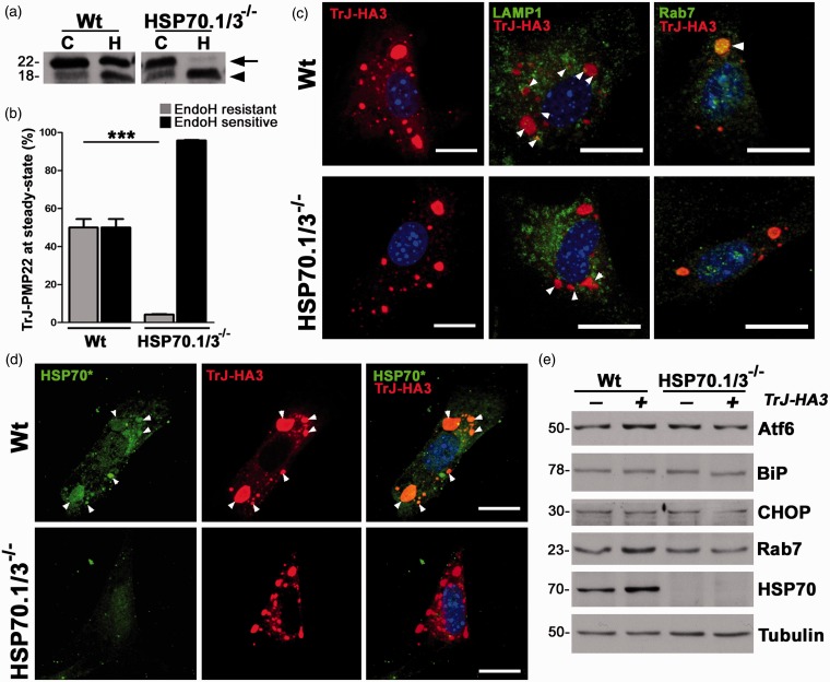
Chaperones, also called heat shock proteins (HSPs), transiently interact with proteins to aid their folding, trafficking, and degradation, thereby directly influencing the transport of newly synthesized molecules. Induction of chaperones provides a potential therapeutic approach for protein misfolding disorders, such as peripheral myelin protein 22 (PMP22)-associated peripheral neuropathies. Cytosolic aggregates of PMP22, linked with a demyelinating Schwann cell phenotype, result in suppression of proteasome activity and activation of proteostatic mechanisms, including the heat shock pathway. Although the beneficial effects of chaperones in preventing the aggregation and improving the trafficking of PMP22 have been repeatedly observed, the requirement for HSP70 in events remains elusive. In this study, we show that activation of the chaperone pathway in fibroblasts from PMP22 duplication-associated Charcot-Marie-Tooth disease type 1A patient with an FDA-approved small molecule increases HSP70 expression and attenuates proteasome dysfunction. Using cells from an HSP70.1/3(-/-) (inducible HSP70) mouse model, we demonstrate that under proteotoxic stress, this chaperone is critical in preventing the aggregation of PMP22, and this effect is aided by macroautophagy. When examined at steady-state, HSP70 appears to play a minor role in the trafficking of wild-type-PMP22, while it is crucial for preventing the buildup of the aggregation-prone Trembler-J-PMP22. HSP70 aids the processing of Trembler-J-PMP22 through the Golgi and its delivery to lysosomes via Rab7-positive vesicles. Together, these results demonstrate a key role for inducible HSP70 in aiding the processing and hindering the accumulation of misfolded PMP22, which in turn alleviates proteotoxicity within the cells.

Keywords: Charcot–Marie–Tooth disease; Trembler-J; chaperones; neuropathy; proteasome.


Figures

Figure 1.
Figure 1.
Activation of the heat shock pathway attenuates proteasome dysfunction in CMT1A patient fibroblasts. (a) Dermal fibroblasts from a control, nonneuropathic individual (Ct) and a CMT1A patient were transfected with Ub-G76V-GFP reporter and treated with either DMSO (−) or BIIB021 (+) and imaged for GFP. The number of GFP-positive cells were counted in fixed microscopic fields (0.1 mm2) and plotted as percentage of total (Hoechst positive) (n = 3 independent experiments). (b) Representative images of Ub-GFP +cells in control and CMT1A cultures after treatment with DMSO or BIIB021. Hoechst dye (blue) was used to visualize nuclei. Scale bar, as shown. (c) The 20S chymotrypsin-like activity of the fibroblasts from the Ct and the CMT1A patient, after treatment with DMSO (−) or BIIB021 (+) is graphed as percentage of DMSO-treated Ct cultures (n = 3 independent experiments). (a, c) Graphs plotted as means ± SEM. Unpaired Student’s t test, *p < .05, **p < .01, ***p < .001, n.s.; nonsignificant. (d) Steady-state levels of HSPs, in fibroblast cell lysates (15 µg/lane) after treatment with DMSO (−) or BIIB021 (+). Tubulin serves as a loading control. Molecular mass on left, in kDa. CMT1A = Charcot–Marie–Tooth disease type 1A; DMSO = dimethyl sulfoxide; HSP = heat shock protein; GFP = green fluorescent protein.
Figure 2.
Figure 2.
HSP70 hinders the aggregation of PMP22 in MEFs. (a) Double immunostaining of Wt and HSP70.1/3−/− MEFs with anti-PMP22 (green) and anti-ubiquitin (red) antibodies, after 16 hr treatment with lactacystin (Lc) or DMSO (insets). Arrows point to PMP22- and Ub-positive protein aggregates. Nuclei are stained with Hoechst dye (blue). Scale bars, 10 µm. (b) Whole cell lysates (15 µg/lane) of Wt and HSP70.1/3−/− MEFs after treatment with DMSO (Ct) or Lc were analyzed for the levels of ubiquitinated substrates and inducible HSP70. Nonconjugated mono-ubiquitin is designated as mUb. GAPDH is shown as a protein loading control. Molecular mass on left, in kDa. (c) The 20S chymotrypsin-like activity of Wt and HSP70.1/3−/− MEFs is graphed as means ± SEM (n = 3 independent cultures). Unpaired Student’s t test, ***p < .001. PMP22 = peripheral myelin protein 22; DMSO = dimethyl sulfoxide; HSP = heat shock protein; MEFs = Mouse embryonic fibroblasts; pUb = poly-ubiquitinated; mUb = mono-ubiquitin; GAPDH = Glyceraldehyde 3-phosphate dehydrogenase.
Figure 3.
Figure 3.
Pharmacological induction of chaperones suppresses PMP22 aggregation in Wt, but not in HSP70.1/3−/− MEFs. (a) Representative images of Wt and HSP70.1/3−/− MEFs after 16 hr treatment with Lc and pretreatment with BIIB021 or DMSO (insets) for 8 hr. Arrows indicate the PMP22- (green) and Ub- (red) positive aggregates. Hoechst dye (blue) is used to visualize the nuclei. Scale bars, 20 µm. (b) The levels of poly-ubiquitinated substrates (pUb) were analyzed in whole cell lysates (15 µg/lane) of Wt and HSP70.1/3−/− MEFs after pretreatment with BIIB021 (+) or DMSO (−), followed by Lc treatment. The nonconjugated mUb is also marked. (c) Whole cell lysates (20 µg/lane) of Wt and HSP70.1/3−/− MEFs, treated with BIIB021 (+) or DMSO (−) for 8 hr were analyzed for the levels of inducible HSP70. (b, c) GAPDH serves as a loading control. Molecular mass in kDa, on left. PMP22 = peripheral myelin protein 22; MEFs = Mouse embryonic fibroblasts; HSP = heat shock protein; DMSO = dimethyl sulfoxide; mUb = mono-ubiquitin; GAPDH = Glyceraldehyde 3-phosphate dehydrogenase.
Figure 4.
Figure 4.
Autophagy aids in the reduction of PMP22 aggregates by HSP70. (a) The percentage of PMP22- and (b) Ub-positive aggregate-containing cells after the indicated treatments were counted and graphed. Data are shown as means ± SEM. Unpaired Student’s t test, ###p < .001, #p < .05 (across genotypes between Lc-treated cultures), *p < .05, **p < .01, ***p < .001, n.s.; nonsignificant (n = 3 independent experiments). (c) PMP22 (green) and Ub (red)-reactive protein aggregates in Wt and HSP70.1/3−/− MEFs after sequential treatment with BIIB021 and Lc alone (insets, BIIB + Lc), or in combination with 3-MA (BIIB021 + Lc + 3MA). Arrows point to PMP22 and Ub-positive aggregates. Asterisks mark cells with dispersed aggregates. Hoechst dye (blue) was used to visualize nuclei. Scale bars, 20 µm. (d) Whole cell lysates (15 µg/lane) of Wt and HSP70.1/3−/− MEFs were analyzed for levels of pUb-substrates and inducible HSP70. Tubulin serves as a protein loading control. Molecular mass in kDa, on left. PMP22 = peripheral myelin protein 22; MEFs = Mouse embryonic fibroblasts; HSP = heat shock protein; pUb = poly-ubiquitinated.
Figure 5.
Figure 5.
Inducible HSP70 is not critical for Wt-PMP22 trafficking. (a) Lysates of sciatic nerves (5 µg/lane) from Wt and HSP70.1/3−/− mice were incubated with no enzyme (Control, C), Endoglycosidase H (EndoH, H), or N-Glycosidase F (PNGaseF, N). (b) Quantification of EndoH-sensitive and EndoH-resistant PMP22 fractions in sciatic nerve lysates from independent experiments (n = 3). (c) Whole cell lysates (35 µg/lane) of Wt and HSP70.1/3−/− MEFs, transfected with Wt-Myc3, were incubated with no enzyme (C), EndoH (H) or PNGaseF (N) and probed for myc. (a, c) Arrows point to the EndoH- resistant fractions while arrowheads mark the EndoH-sensitive fractions. Molecular mass in kDa, on left. (d) Quantification of EndoH-sensitive and EndoH-resistant Wt-Myc3 fractions from independent experiments (n = 3). (b, d) Graphs plotted as means ± SEM. Unpaired Student’s t test, n.s. = nonsignificant. (e) MEFs transfected with Wt-Myc3 were immunostained for myc (red) and Concanavalin A (ConA, ER marker; green). Arrows point at the cell boundary. Hoechst dye (blue) was used to visualize the nuclei. Scale bar, as shown. PMP22 = peripheral myelin protein 22; HSP = heat shock protein; MEFs = Mouse embryonic fibroblasts.
Figure 6.
Figure 6.
Absence of HSP70 severely impairs the processing of TrJ-PMP22. (a) Lysates from Wt and HSP70.1/3−/− MEFs (35 µg/lane), transfected with TrJ-HA3, were treated with either no enzyme (C) or EndoH (H). Membranes were probed with an anti-HA antibody. Arrow points to the EndoH-resistant protein fractions while arrowhead marks the EndoH-sensitive form. (b) Quantification of the EndoH-resistant and EndoH-sensitive TrJ-HA3 fractions from (a) from independent experiments (n = 3). Graph plotted as means ± SEM. Unpaired Student’s t test, ***p < .001. (c) Wt (upper panel) and HSP70.1/3−/− MEFs (lower panel), transfected with TrJ-HA3, were immunostained for either HA (red) alone or in combination with LAMP1 (green) or Rab7 (green). (d) TrJ-PMP22 expressing Wt and HSP70.1/3−/− MEFs were probed with anti- HA (red) and anti-HSP70 antibodies. The anti-HSP70 antibody recognizes both the inducible and constitutive forms of HSP70 (HSP70*, green). (c, d) Hoechst dye (blue) was used to visualize the nuclei. Scale bars, 20 µm. Arrowheads mark areas of colocalization. (e) Lysates (30 µg/lane) of transfected MEFs were probed for the indicated proteins. Tubulin serves as a protein loading control. (a, e) Molecular mass in kDa, on left. PMP22 = peripheral myelin protein 22; HSP = heat shock protein; MEFs = Mouse embryonic fibroblasts; TrJ = Trembler-J; CHOP = C/EBP homologous protein; BiP = Binding immunoglobulin protein.
Figure 7.
Figure 7.
Working model of Wt and TrJ-PMP22 trafficking in the presence and absence of HSP70. (a) In normal MEFs, Wt-PMP22 and TrJ-PMP22 are trafficked out the ER (a). ‘X’ (red) represents unknown protein/s involved in the translocation. Approximately 22% of Wt-PMP22 and about half of TrJ-PMP22 are translocated (b) to the proteasome (c) for degradation. Intracellular PMP22 aggregates impair the proteasome function (d). However, the presence of HSP70 hinders aggregate formation, by increasing the solubility of cytoplasmic PMP22 (e) and facilitating the removal of misfolded protein through autophagy (f). Aggregates engulfed by autophagosomes are degraded in LAMP1-positive lysosomes (g). Fractions of Wt- and TrJ-PMP22 are processed through the Golgi (i) and inserted into the plasma membrane (j). Due to structural instability, the TrJ-PMP22 is endocytosed (k) via Rab7-positive endosomes for degradation (l) by lysosomes. (b) In case of HSP70.1/3−/− cells, there is increased protein aggregate formation that can impair the proteasome function (d). In HSP70-deficient cells, Wt-PMP22 still reaches the plasma membrane (i, j), while TrJ-PMP22 fails to reach the Golgi in appreciable levels. PMP22 = peripheral myelin protein 22; HSP = heat shock protein; TrJ = Trembler-J; MEFs = Mouse embryonic fibroblasts.

Similar articles

Cited by

References

    1. Baechner D., Liehr T., Hameister H., Altenberger H., Grehl H., Suter U., Rautenstrauss B. (1995) Widespread expression of the peripheral myelin protein-22 gene (PMP22) in neural and non-neural tissues during murine development. Journal of Neuroscience Research 42: 733–741. - PubMed
    1. Bennett E. J., Bence N. F., Jayakumar R., Kopito R. R. (2005) Global impairment of the ubiquitin-proteasome system by nuclear or cytoplasmic protein aggregates precedes inclusion body formation. Molecular Cell 17: 351–365. - PubMed
    1. Bobkova N. V., Garbuz D. G., Nesterova I., Medvinskaya N., Samokhin A., Alexandrova I., Evgen'ev M. (2014) Therapeutic effect of exogenous hsp70 in mouse models of Alzheimer's disease. Journal of Alzheimer’s Disease 38: 425–435. - PubMed
    1. Bucci C., Thomsen P., Nicoziani P., McCarthy J., van Deurs B. (2000) Rab7: A key to lysosome biogenesis. Molecular Biology of the Cell 11: 467–480. - PMC - PubMed
    1. Dantuma N. P., Lindsten K., Glas R., Jellne M., Masucci M. G. (2000) Short-lived green fluorescent proteins for quantifying ubiquitin/proteasome-dependent proteolysis in living cells. Nature Biotechnology 18: 538–543. - PubMed

Publication types

MeSH terms

LinkOut - more resources