

Myelin abnormalities in the optic and sciatic nerves in mice with GM1-gangliosidosis
- PMID: 25694553
- PMCID: PMC4342369
- DOI: 10.1177/1759091415568913
Myelin abnormalities in the optic and sciatic nerves in mice with GM1-gangliosidosis
Abstract
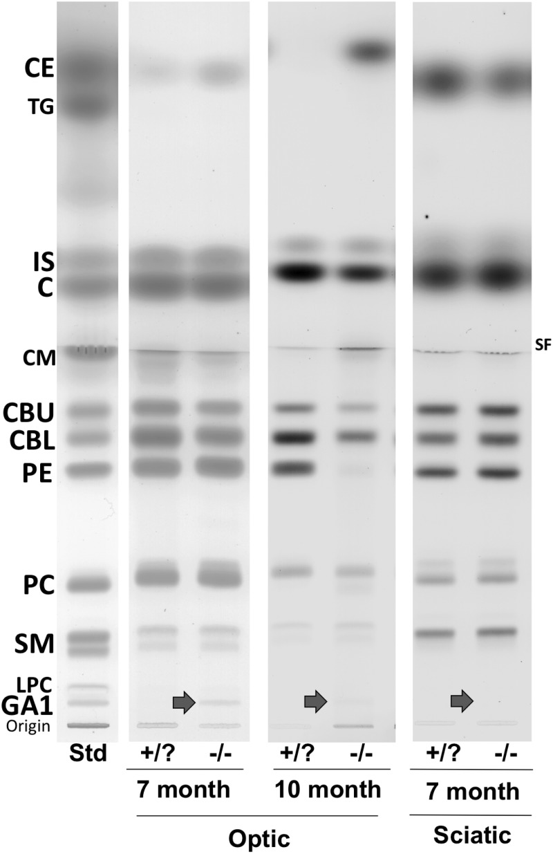
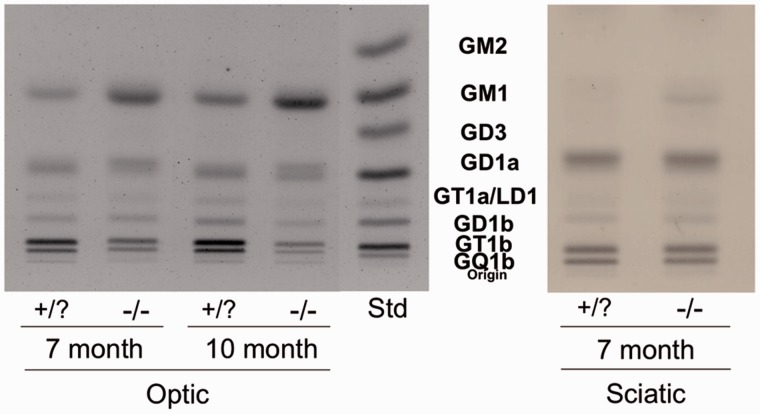
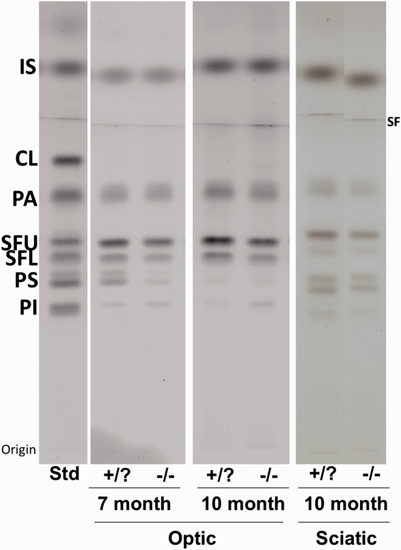
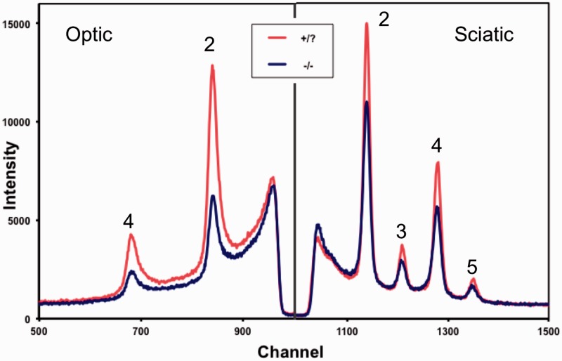
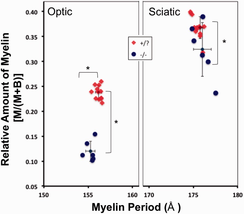
GM1-gangliosidosis is a glycosphingolipid lysosomal storage disease involving accumulation of GM1 and its asialo form (GA1) primarily in the brain. Thin-layer chromatography and X-ray diffraction were used to analyze the lipid content/composition and the myelin structure of the optic and sciatic nerves from 7- and 10-month old β-galactosidase (β-gal) +/? and β-gal -/- mice, a model of GM1gangliosidosis. Optic nerve weight was lower in the β-gal -/- mice than in unaffected β-gal +/? mice, but no difference was seen in sciatic nerve weight. The levels of GM1 and GA1 were significantly increased in both the optic nerve and sciatic nerve of the β-gal -/- mice. The content of myelin-enriched cerebrosides, sulfatides, and plasmalogen ethanolamines was significantly lower in optic nerve of β-gal -/- mice than in β-gal +/? mice; however, cholesteryl esters were enriched in the β-gal -/- mice. No major abnormalities in these lipids were detected in the sciatic nerve of the β-gal -/- mice. The abnormalities in GM1 and myelin lipids in optic nerve of β-gal -/- mice correlated with a reduction in the relative amount of myelin and periodicity in fresh nerve. By contrast, the relative amount of myelin and periodicity in the sciatic nerves from control and β-gal -/- mice were indistinguishable, suggesting minimal pathological involvement in sciatic nerve. Our results indicate that the greater neurochemical pathology observed in the optic nerve than in the sciatic nerve of β-gal -/- mice is likely due to the greater glycolipid storage in optic nerve.
Keywords: X-ray diffraction; cerebrosides; gangliosides; lipids.
© The Author(s) 2015.
Figures
Similar articles
-
N-butyldeoxygalactonojirimycin reduces neonatal brain ganglioside content in a mouse model of GM1 gangliosidosis.J Neurochem. 2004 May;89(3):645-53. doi: 10.1046/j.1471-4159.2004.02381.x. J Neurochem. 2004. PMID: 15086521
-
Inheritance of lysosomal acid beta-galactosidase activity and gangliosides in crosses of DBA/2J and knockout mice.Biochem Genet. 2004 Aug;42(7-8):241-57. doi: 10.1023/b:bigi.0000034429.55418.71. Biochem Genet. 2004. PMID: 15487588
-
GM1-gangliosidosis in American black bears: clinical, pathological, biochemical and molecular genetic characterization.Mol Genet Metab. 2014 Apr;111(4):513-21. doi: 10.1016/j.ymgme.2014.02.002. Epub 2014 Feb 13. Mol Genet Metab. 2014. PMID: 24581871
-
[Structural basis for β-galactosidase associated with lysosomal disease].Yakugaku Zasshi. 2013;133(5):509-17. doi: 10.1248/yakushi.13-00001-1. Yakugaku Zasshi. 2013. PMID: 23649392 Review. Japanese.
-
[beta-galactosidosis--GM1 gangliosidosis and Morquio B disease].Nihon Rinsho. 1995 Dec;53(12):2960-6. Nihon Rinsho. 1995. PMID: 8577043 Review. Japanese.
Cited by
-
Adeno-associated virus expressing a blood-brain barrier-penetrating enzyme improves GM1 gangliosidosis in a preclinical model.J Clin Invest. 2025 Apr 8;135(12):e180724. doi: 10.1172/JCI180724. eCollection 2025 Jun 16. J Clin Invest. 2025. PMID: 40198143 Free PMC article.
-
GM1 Gangliosidosis: Mechanisms and Management.Appl Clin Genet. 2021 Apr 9;14:209-233. doi: 10.2147/TACG.S206076. eCollection 2021. Appl Clin Genet. 2021. PMID: 33859490 Free PMC article. Review.
-
Axonopathy and Reduction of Membrane Resistance: Key Features in a New Murine Model of Human GM1-Gangliosidosis.J Clin Med. 2020 Apr 2;9(4):1004. doi: 10.3390/jcm9041004. J Clin Med. 2020. PMID: 32252429 Free PMC article.
-
Ganglioside GM1 and the Central Nervous System.Int J Mol Sci. 2023 May 31;24(11):9558. doi: 10.3390/ijms24119558. Int J Mol Sci. 2023. PMID: 37298512 Free PMC article. Review.
-
White Matter Pathology as a Barrier to Gangliosidosis Gene Therapy.Front Cell Neurosci. 2021 Aug 12;15:682106. doi: 10.3389/fncel.2021.682106. eCollection 2021. Front Cell Neurosci. 2021. PMID: 34456684 Free PMC article. Review.
References
-
- Agrawal D., Hawk R., Avila R. L., Inouye H., Kirschner D. A. (2009) Internodal myelination during development quantitated using X-ray diffraction. Journal of Structural Biology 168: 521–526. - PubMed
-
- Ando S., Chang N. C., Yu R. K. (1978) High-performance thin-layer chromatography and densitometric determination of brain ganglioside compositions of several species. Analytical Biochemistry 89: 437–450. - PubMed
-
- Ando S., Tanaka Y., Toyoda Y., Kon K. (2003) Turnover of myelin lipids in aging brain. Neurochemical Research 28: 5–13. - PubMed
Publication types
MeSH terms
Substances
Grants and funding
LinkOut - more resources
Full Text Sources
Other Literature Sources
