. 2015 May;89(9):4942-50.
doi: 10.1128/JVI.03688-14. Epub 2015 Feb 18.

Broad-spectrum inhibitors against 3C-like proteases of feline coronaviruses and feline caliciviruses

Affiliations

Broad-spectrum inhibitors against 3C-like proteases of feline coronaviruses and feline caliciviruses

Yunjeong Kim et al. J Virol. 2015 May.

Abstract

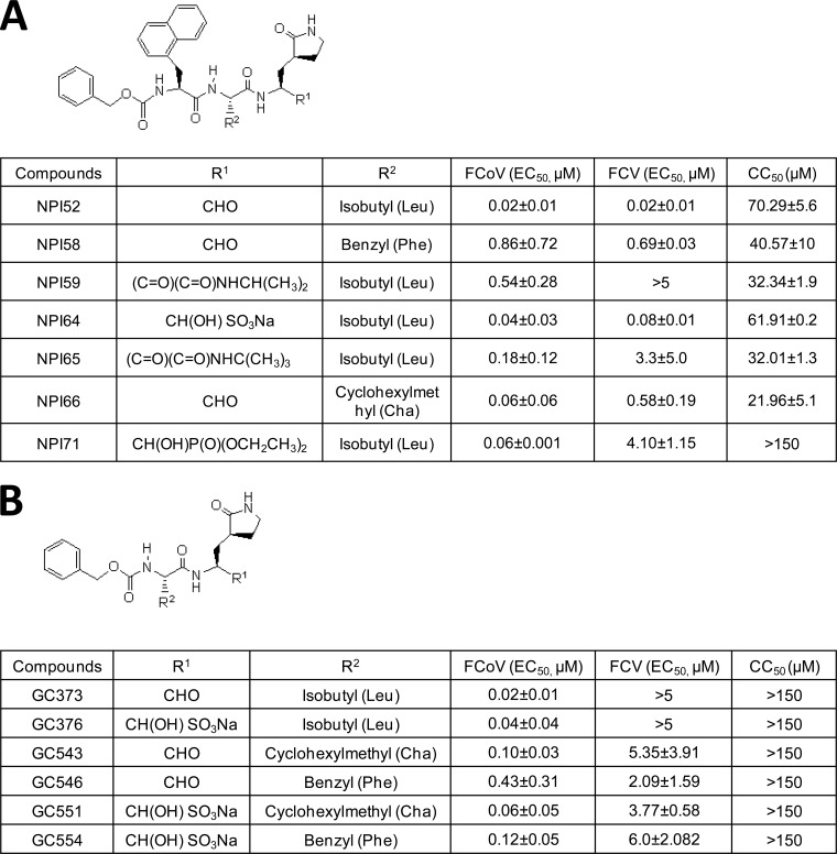
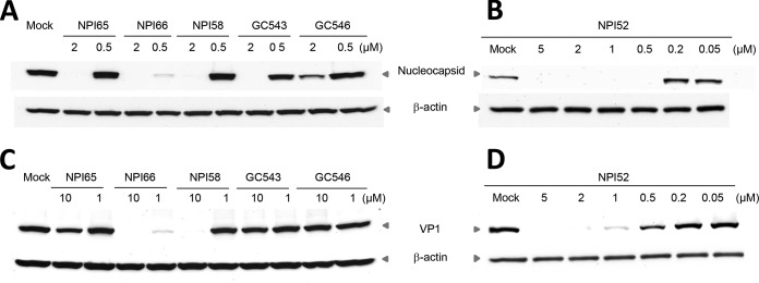
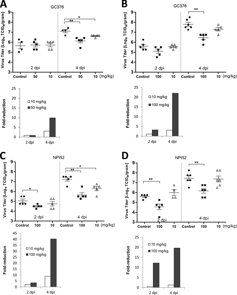
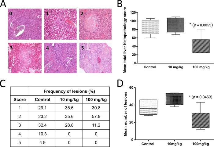
Feline infectious peritonitis and virulent, systemic calicivirus infection are caused by certain types of feline coronaviruses (FCoVs) and feline caliciviruses (FCVs), respectively, and are important infectious diseases with high fatality rates in members of the Felidae family. While FCoV and FCV belong to two distinct virus families, the Coronaviridae and the Caliciviridae, respectively, they share a dependence on viral 3C-like protease (3CLpro) for their replication. Since 3CLpro is functionally and structurally conserved among these viruses and essential for viral replication, 3CLpro is considered a potential target for the design of antiviral drugs with broad-spectrum activities against these distinct and highly important viral infections. However, small-molecule inhibitors against the 3CLpro enzymes of FCoV and FCV have not been previously identified. In this study, derivatives of peptidyl compounds targeting 3CLpro were synthesized and evaluated for their activities against FCoV and FCV. The structures of compounds that showed potent dual antiviral activities with a wide margin of safety were identified and are discussed. Furthermore, the in vivo efficacy of 3CLpro inhibitors was evaluated using a mouse model of coronavirus infection. Intraperitoneal administration of two 3CLpro inhibitors in mice infected with murine hepatitis virus A59, a hepatotropic coronavirus, resulted in significant reductions in virus titers and pathological lesions in the liver compared to the findings for the controls. These results suggest that the series of 3CLpro inhibitors described here may have the potential to be further developed as therapeutic agents against these important viruses in domestic and wild cats. This study provides important insights into the structure and function relationships of 3CLpro for the design of antiviral drugs with broader antiviral activities.

Importance: Feline infectious peritonitis virus (FIPV) is the leading cause of death in young cats, and virulent, systemic feline calicivirus (vs-FCV) causes a highly fatal disease in cats for which no preventive or therapeutic measure is available. The genomes of these distinct viruses, which belong to different virus families, encode a structurally and functionally conserved 3C-like protease (3CLpro) which is a potential target for broad-spectrum antiviral drug development. However, no studies have previously reported a structural platform for the design of antiviral drugs with activities against these viruses or on the efficacy of 3CLpro inhibitors against coronavirus infection in experimental animals. In this study, we explored the structure-activity relationships of the derivatives of 3CLpro inhibitors and identified inhibitors with potent dual activities against these viruses. In addition, the efficacy of the 3CLpro inhibitors was demonstrated in mice infected with a murine coronavirus. Overall, our study provides the first insight into a structural platform for anti-FIPV and anti-FCV drug development.


Figures

FIG 1
FIG 1
Chemical structures of tripeptidyl (A) and dipeptidyl (B) compounds and the mean and standard error of the means (SEM) of the EC50 of the compounds against FCoV or FCV. Each compound was added to CRFK cells, and the cells were immediately infected with FCoV WSU-79-1146 or FCV Urbana. Cells were further incubated in the presence of each compound for up to 24 h. Virus titers were determined using the TCID50 method, and the EC50 were calculated. Compound cytotoxicity (CC50) was measured after incubating the cells with each compound for 24 h.
FIG 2
FIG 2
Western blot analysis of the effects of the compounds on expression of the FCoV nucleocapsid protein or FCV VP1 in CRFK cells. Cells were mock treated or treated with each compound and immediately infected with FCoV WSU-79-1146 or FCV Urbana. The cells were then further incubated for 12 h. Cell lysates were prepared and analyzed for expression of viral proteins on Western blots. β-Actin was used as a loading control.
FIG 3
FIG 3
Multiple-sequence alignments of 3CLpro enzymes from FCV (A) and FCoV and MHV A59 (C) and ribbon presentations of three-dimensional structural models for FCV 3CLpro (B) and FCoV 3CLpro (D). (A and C) The catalytic residues E60, C122, and H39 of FCV 3CLpro (A) and H41 and C144 of FCoV 3CLpro and MHV A59 3CLpro are indicated by red arrowheads (C). (B and D) The structure model of FCoV 3CLpro was built by use of the EasyModeller (version 4.0) program (29) and the 3CLpro structure of TGEV (Protein Data Bank accession number 2AMP) as the template. The structural model of FCV 3CLpro was built by use of the EasyModeller program and the 3Cpro enzymes of rhinovirus, poliovirus, and hepatitis A virus and the 3CLpro of human norovirus (Protein Data Bank accession numbers 1CQQ, 1L1N, 1QA7, and 2LNC, respectively) (30) as the templates. The amino acids in the catalytic triad (E60, C122, and H39 for FCV 3CLpro) (B) and dyad (H41 and C144 for FCoV 3CLpro) (C) are shown in the blue boxes.
FIG 4
FIG 4
Effects of 3CLpro inhibitor treatment on MHV A59 titers. Four- to 5-week-old BALB/c mice were intraperitoneally inoculated with MHV A59 at 5.2 × 10⁵ (A) or 7.2 × 10⁴ (B to D) TCID50/mouse and treated with drug vehicle, GC376 (10, 50, or 100 mg/kg/day), or NPI52 (10 or 100 mg/kg/day) divided into two doses per day starting at 4 h prior to virus infection. Scatter plots show the mean ± standard error of the mean virus titers in the livers of mice receiving mock treatment (drug vehicle) or treatment with GC376 (A and B) or NPI52 (C and D) at 2 or 4 days after virus infection (dpi, days postinfection). Virus titers are expressed as log10 TCID50 per gram of liver tissue. Bar graphs show the fold reduction of the geometric mean virus titers in the treatment groups compared to the titers in the control group. Asterisks indicate significant differences between the control and the treated groups (*, P < 0.05; **, P < 0.01).
FIG 5
FIG 5
Histopathology changes in the livers of mice treated with NPI52. (A) Panels 0 through 5 show microscopic lesions for increasing histopathology severity scores of 0 through 5, respectively. Score 0, minimal change; scores 1 and 2, multifocal areas of necrosis; and scores 3 to 5, coalescing areas of necrosis. (B) A box-and-whisker plot showing the mean total liver histopathology score for each group. (C) A table showing the frequency of histopathology scores in four liver samples per group (for the group treated with NPI52 at 10 mg/kg) or five liver samples per group (for the control group and the group treated with NPI52 at 100 mg/kg). (D) A box-and-whisker plot showing the mean number of lesions per mouse liver for each group. Asterisks indicate statistically significant differences between control mice and mice treated with NPI52 at 100 mg/kg (P < 0.05). The whiskers represent the 5% and 95% confidence intervals, and the boxes represent the 25% and 75% confidence intervals. The lines in the middle of the boxes represent the medians for the data.

Similar articles

Cited by

References

    1. Rohrbach BW, Legendre AM, Baldwin CA, Lein DH, Reed WM, Wilson RB. 2001. Epidemiology of feline infectious peritonitis among cats examined at veterinary medical teaching hospitals. J Am Vet Med Assoc 218:1111–1115. doi:10.2460/javma.2001.218.1111. - DOI - PubMed
    1. Foley JE, Poland A, Carlson J, Pedersen NC. 1997. Risk factors for feline infectious peritonitis among cats in multiple-cat environments with endemic feline enteric coronavirus. J Am Vet Med Assoc 210:1313–1318. - PubMed
    1. Pedersen NC. 2009. A review of feline infectious peritonitis virus infection: 1963–2008. J Feline Med Surg 11:225–258. doi:10.1016/j.jfms.2008.09.008. - DOI - PMC - PubMed
    1. Benetka V, Kubber-Heiss A, Kolodziejek J, Nowotny N, Hofmann-Parisot M, Mostl K. 2004. Prevalence of feline coronavirus types I and II in cats with histopathologically verified feline infectious peritonitis. Vet Microbiol 99:31–42. doi:10.1016/j.vetmic.2003.07.010. - DOI - PMC - PubMed
    1. Pedersen NC, Black JW, Boyle JF, Evermann JF, McKeirnan AJ, Ott RL. 1984. Pathogenic differences between various feline coronavirus isolates. Adv Exp Med Biol 173:365–380. doi:10.1007/978-1-4615-9373-7_36. - DOI - PubMed

Publication types

MeSH terms

LinkOut - more resources