. 2015 May;89(9):4907-17.
doi: 10.1128/JVI.00338-15. Epub 2015 Feb 18.

Murine coronavirus ubiquitin-like domain is important for papain-like protease stability and viral pathogenesis

Affiliations

Murine coronavirus ubiquitin-like domain is important for papain-like protease stability and viral pathogenesis

Anna M Mielech et al. J Virol. 2015 May.

Abstract

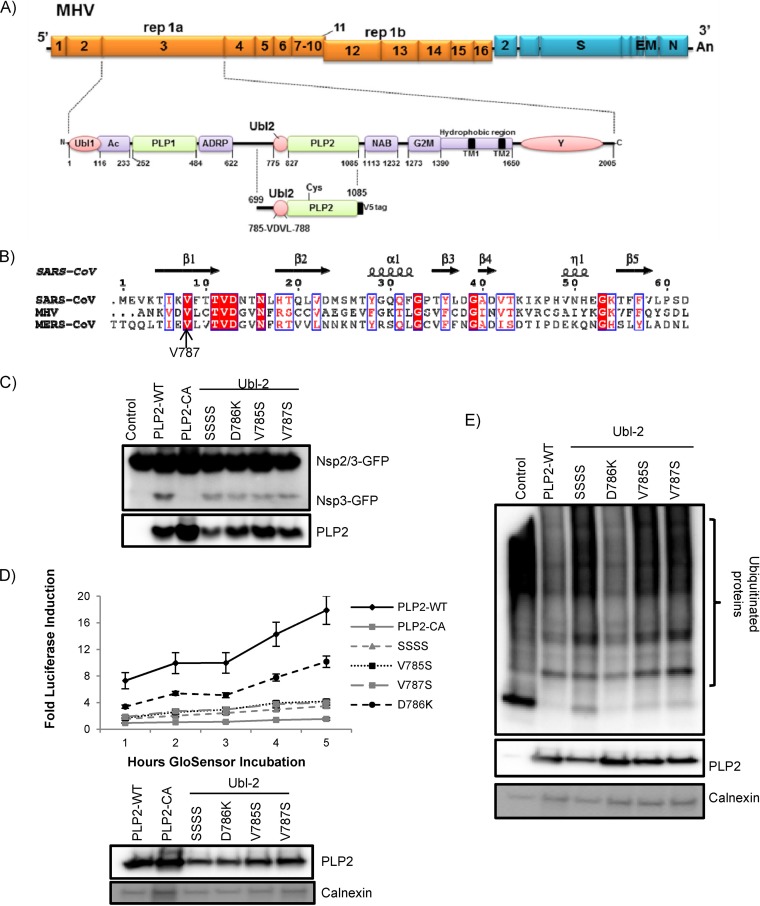
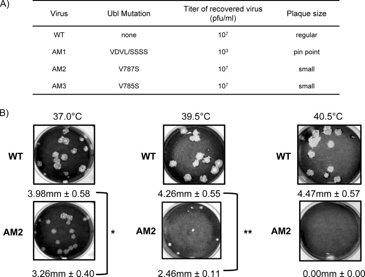
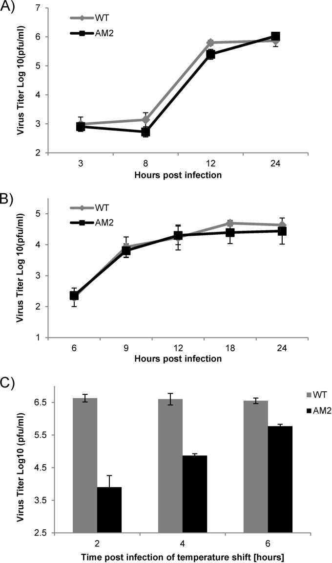
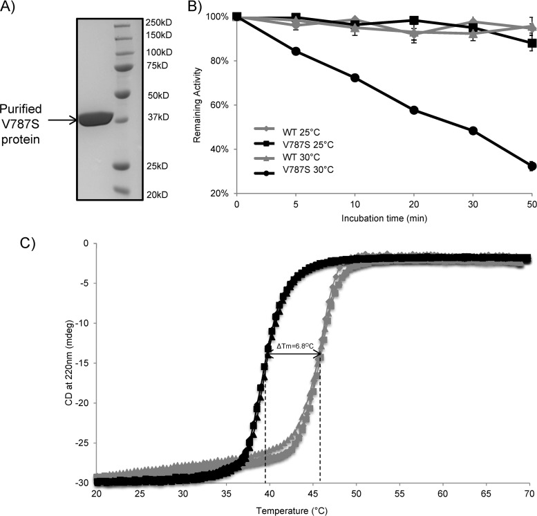
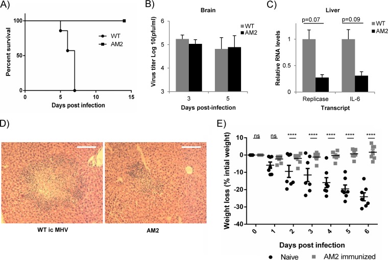
Ubiquitin-like domains (Ubls) now are recognized as common elements adjacent to viral and cellular proteases; however, their function is unclear. Structural studies of the papain-like protease (PLP) domains of coronaviruses (CoVs) revealed an adjacent Ubl domain in severe acute respiratory syndrome CoV, Middle East respiratory syndrome CoV, and the murine CoV, mouse hepatitis virus (MHV). Here, we tested the effect of altering the Ubl adjacent to PLP2 of MHV on enzyme activity, viral replication, and pathogenesis. Using deletion and substitution approaches, we identified sites within the Ubl domain, residues 785 to 787 of nonstructural protein 3, which negatively affect protease activity, and valine residues 785 and 787, which negatively affect deubiquitinating activity. Using reverse genetics, we engineered Ubl mutant viruses and found that AM2 (V787S) and AM3 (V785S) viruses replicate efficiently at 37°C but generate smaller plaques than wild-type (WT) virus, and AM2 is defective for replication at higher temperatures. To evaluate the effect of the mutation on protease activity, we purified WT and Ubl mutant PLP2 and found that the proteases exhibit similar specific activities at 25°C. However, the thermal stability of the Ubl mutant PLP2 was significantly reduced at 30°C, thereby reducing the total enzymatic activity. To determine if the destabilizing mutation affects viral pathogenesis, we infected C57BL/6 mice with WT or AM2 virus and found that the mutant virus is highly attenuated, yet it replicates sufficiently to elicit protective immunity. These studies revealed that modulating the Ubl domain adjacent to the PLP reduces protease stability and viral pathogenesis, revealing a novel approach to coronavirus attenuation.

Importance: Introducing mutations into a protein or virus can have either direct or indirect effects on function. We asked if changes in the Ubl domain, a conserved domain adjacent to the coronavirus papain-like protease, altered the viral protease activity or affected viral replication or pathogenesis. Our studies using purified wild-type and Ubl mutant proteases revealed that mutations in the viral Ubl domain destabilize and inactivate the adjacent viral protease. Furthermore, we show that a CoV encoding the mutant Ubl domain is unable to replicate at high temperature or cause lethal disease in mice. Our results identify the coronavirus Ubl domain as a novel modulator of viral protease stability and reveal manipulating the Ubl domain as a new approach for attenuating coronavirus replication and pathogenesis.


Figures

FIG 1
FIG 1
MHV-A59 Ubl-2 domain influences papain-like protease activity. (A) Schematic diagram of MHV-A59 ORFs and the Ubl-2 and papain-like protease (PLP) domains within nonstructural protein 3. Expression plasmid PLP2 and the VDVL region (aa 785 to 788) are indicated. (B) Alignment of the predicted ubiquitin-like domains from MHV and MERS-CoV with the structural information from the Ubl domain of SARS-CoV (PDB code 2FE8). (C) HEK293T cells were transfected with plasmids expressing the PLP2 wild type, PLP2 catalytic mutant CA, and VDV Ubl-2 domain mutants in the presence of plasmid expressing the nsp2/3-GFP substrate. At 24 h posttransfection, cells were lysed and analyzed by Western blotting. (D) PLP2 activity in live-cell assay. HEK293T cells were transfected with pGlo-RLKGG and the indicated PLP2 expression plasmids. At 14 h postinfection, GloSensor was added and luminescence was assessed hourly. (E) HEK293T cells were transfected with Flag-Ub expression plasmid and the wild type (PLP2-WT) or indicated Ubl-2 mutants. Cells were lysed 24 h posttransfection and analyzed by Western blotting. The figure shows representative data from at least two independent experiments.
FIG 2
FIG 2
Murine coronavirus AM2 is temperature sensitive for replication. (A) Summary of the characteristics of Ubl mutant viruses recovered by reverse genetics. (B) Representative plaques generated by wild-type MHV (WT) or AM2 during incubation at the indicated temperature. DBT cells were inoculated with either the WT or AM2, incubated at the indicated temperature, and fixed and stained 48 h postinfection. The diameter of each plaque was measured, and average diameters and standard deviations are indicated for each virus. *, P < 0.05; **, P < 0.001. Representative data from at least two independent experiments are shown.
FIG 3
FIG 3
Replication kinetics of AM2. (A) AM2 replication is similar to that of MHV-WT at 37°C. DBT cells were infected with AM2 or the WT at an MOI of 0.1. (B) Bone marrow-derived macrophages were infected with AM2 or the WT at an MOI of 1. At the indicated time points, supernatant was harvested and virus titer determined by plaque assay at 37°C. (C) Temperature shift reduces yield of AM2. DBT cells were infected at an MOI of 0.1 at 37°C with AM2 or the WT. At the indicated time point postinfection, cells were moved to 39.5°C until 14 hpi, when supernatant was harvested and virus titer was determined by plaque assay at 37°C. The error bars represent standard deviations within single experiments. Shown are representative data from at least two independent experiments.
FIG 4
FIG 4
Ubl-2 domain is important for MHV PLP2 thermal stability. (A) SDS-PAGE analysis of the final purified PLP2-V787S enzyme. Wild-type and PLP2-V787S mutant PLP2 were expressed and purified from E. coli as described in Materials and Methods. (B) Temperature-dependent inactivation of wild-type PLP2 and PLP2-V787S mutant. The specific activities of the WT and the V787S mutant PLP2 enzymes were measured after incubation at 25°C and 30°C for different time periods. The specific activities then were normalized to the activity at 0 min (Ratet/Rate0 indicates the rate at time t over the initial rate). Kinetic data for the V787S mutant incubated at 30°C were fit to a first-order exponential decay model (Ratet/Rate0 = ekt). The calculated inactivation rate constant (kinact) for the V787S mutant at 30°C is 0.025 ± 0.001 min−1, and the half-life (t1/2) is 27.7 ± 1.1 min. (C) CD melting curves of WT and PLP2-V787S mutant PLP2. The thermal stability of WT and V787S mutant PLP2 was determined by measuring the CD signal at 220 nm as a function of temperature at a step interval of 0.4°C and at a rate of 0.5°C/min. Three independent experiments were performed for both WT PLP2 (gray) and the V787S mutant (black).
FIG 5
FIG 5
MHV AM2 is attenuated and generated protective immunity in mice. (A) C57BL/6 mice were infected with 600 PFU WT icMHV or AM2 mutant intracranially and monitored for survival over time (n = 7 for each group). (B) Virus titers in brains of mice infected with 600 PFU WT icMHV or AM2 were determined at the indicated time points p.i. by plaque assay (n = 4 to 5). Error bars represent standard deviations. (C) RNA levels for viral replicase gene and cellular IL-6 were determined using qRT-PCR in livers at day 5 postinfection (n = 6). RNA levels were normalized to actin, and the amount of transcript in WT-infected mice was set to 1. Error bars represent standard errors of the means (SEM). Statistical analysis performed using Student's t test. (D) Representative samples of hematoxylin and eosin staining of formalin-fixed liver samples at day 5 postinfection from mice infected with WT icMHV or AM2 (n = 6 for each group; magnification, ×10). (E) C57BL/6 mice immunized with AM2 mutant and naive age-matched controls were challenged with 6,000 PFU icMHV intracranially 9 weeks after primary infection. The mice were monitored for body weight loss. Error bars represent SEM. ****, P < 0.0001 by two-way analysis of variance (ANOVA). ns, not significant.
FIG 6
FIG 6
X-ray structures of the MHV and SARS-CoV Ubl-2 domains. A superposition of the Ubl-2 domains from MHV PLP2 (blue) and SARS-CoV PLpro (orange; PDB code 2FE8) is shown as a stereo view and resulted in an root mean square deviation of 0.74 Å. Side chains of MHV Ubl-2 residues V785 and V787 and their corresponding residues in SARS-CoV are shown as sticks.

Similar articles

Cited by

References

    1. Peiris JS, Guan Y, Yuen KY. 2004. Severe acute respiratory syndrome. Nat Med 10:S88–S97. doi:10.1038/nm1143. - DOI - PMC - PubMed
    1. Zaki AM, van Boheemen S, Bestebroer TM, Osterhaus AD, Fouchier RA. 2012. Isolation of a novel coronavirus from a man with pneumonia in Saudi Arabia. N Engl J Med 367:1814–1820. doi:10.1056/NEJMoa1211721. - DOI - PubMed
    1. Lau SK, Woo PC, Li KS, Huang Y, Tsoi HW, Wong BH, Wong SS, Leung SY, Chan KH, Yuen KY. 2005. Severe acute respiratory syndrome coronavirus-like virus in Chinese horseshoe bats. Proc Natl Acad Sci U S A 102:14040–14045. doi:10.1073/pnas.0506735102. - DOI - PMC - PubMed
    1. Poon LL, Chu DK, Chan KH, Wong OK, Ellis TM, Leung YH, Lau SK, Woo PC, Suen KY, Yuen KY, Guan Y, Peiris JS. 2005. Identification of a novel coronavirus in bats. J Virol 79:2001–2009. doi:10.1128/JVI.79.4.2001-2009.2005. - DOI - PMC - PubMed
    1. Perlman S, Netland J. 2009. Coronaviruses post-SARS: update on replication and pathogenesis. Nat Rev 7:439–450. doi:10.1038/nrmicro2147. - DOI - PMC - PubMed

Publication types

MeSH terms

Associated data