. 2015 May;89(9):4770-85.
doi: 10.1128/JVI.00097-15. Epub 2015 Feb 18.

CCCTC-binding factor recruitment to the early region of the human papillomavirus 18 genome regulates viral oncogene expression

Affiliations

CCCTC-binding factor recruitment to the early region of the human papillomavirus 18 genome regulates viral oncogene expression

Christian Paris et al. J Virol. 2015 May.

Abstract

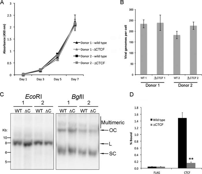
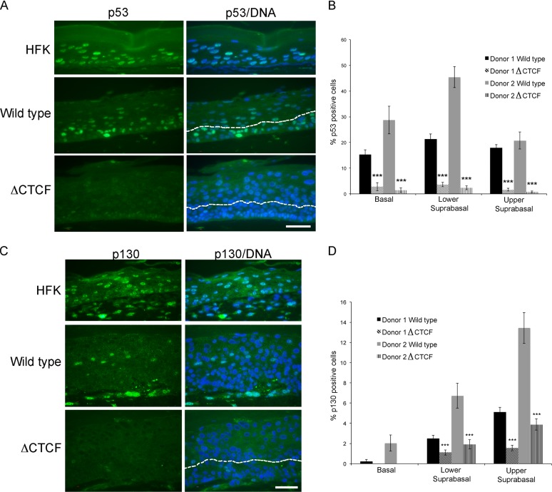
Host cell differentiation-dependent regulation of human papillomavirus (HPV) gene expression is required for productive infection. The host cell CCCTC-binding factor (CTCF) functions in genome-wide chromatin organization and gene regulation. We have identified a conserved CTCF binding site in the E2 open reading frame of high-risk HPV types. Using organotypic raft cultures of primary human keratinocytes containing high-risk HPV18 genomes, we show that CTCF recruitment to this conserved site regulates viral gene expression in differentiating epithelia. Mutation of the CTCF binding site increases the expression of the viral oncoproteins E6 and E7 and promotes host cell proliferation. Loss of CTCF binding results in a reduction of a specific alternatively spliced transcript expressed from the early gene region concomitant with an increase in the abundance of unspliced early transcripts. We conclude that high-risk HPV types have evolved to recruit CTCF to the early gene region to control the balance and complexity of splicing events that regulate viral oncoprotein expression.

Importance: The establishment and maintenance of HPV infection in undifferentiated basal cells of the squamous epithelia requires the activation of a subset of viral genes, termed early genes. The differentiation of infected cells initiates the expression of the late viral transcripts, allowing completion of the virus life cycle. This tightly controlled balance of differentiation-dependent viral gene expression allows the virus to stimulate cellular proliferation to support viral genome replication with minimal activation of the host immune response, promoting virus productivity. Alternative splicing of viral mRNAs further increases the complexity of viral gene expression. In this study, we show that the essential host cell protein CTCF, which functions in genome-wide chromatin organization and gene regulation, is recruited to the HPV genome and plays an essential role in the regulation of early viral gene expression and transcript processing. These data highlight a novel virus-host interaction important for HPV pathogenicity.


Figures

FIG 1
FIG 1
In vitro analysis of the association of CTCF with HPV genomes. (A) Western blot analysis of in vitro-translated CTCF protein. Lysate from ID13 (mouse) cells known to express CTCF was loaded as a positive control alongside wheat germ extract (WGE) and WGE used to translate luciferase (Luc) or CTCF protein. A band running at approximately 140 kDa was present in the ID13 cell lysate, and a slightly smaller band was present in the in vitro-translated CTCF reaction. Human CTCF is an 82-kDa protein but runs at approximately 130 kDa on SDS-PAGE (71), whereas the mouse homologue is slightly larger. (B) An example of an EMSA of CTCF binding to predicted BPV DNA fragments. DNA fragments were amplified and labeled with FAM by PCR. Fragments were mixed with binding buffer only (DNA), in vitro-translated luciferase protein (Luc) (−), or in vitro-translated CTCF protein (+), and protein-DNA complexes were separated on a native acrylamide gel. Free DNA is indicated at the bottom of the gel and protein-DNA complexes near the top. Each fragment was tested a minimum of three times, and the combined results are shown in Table 2. Fragments from the c-Myc locus (positive control), a region of the BPV-1 genome that is known not to bind CTCF (negative control), and fragment 11 from HPV18 and fragments 1 and 10 from HPV31 are shown in the representative EMSA. 18_11 and 31_10 bound CTCF with medium strength (50 to 75% binding compared to the c-Myc positive control), and 31_1 did not bind CTCF in vitro.
FIG 2
FIG 2
Summary of in silico-predicted CTCF binding sites and in vitro analysis. Graphical representations of the HPV16, HPV18, HPV31, HPV11, and HPV6b genomes are shown. ORFs are indicated on each genome (light gray). Predicted CTCF binding sites are represented by the black bars. The hashed bars on the periphery of each genome highlight fragments tested by EMSA, and the dark gray bars on each genome indicate those fragments that bound CTCF in vitro.
FIG 3
FIG 3
Association of CTCF with HPV genomes. Chromatin extracted from HPV16-positive W12 cells (A) and HPV18-positive HFKs (B) was immunoprecipitated with control antibody (rabbit IgG for W12 and FLAG M2 antibody for HPV18 HFKs) or CTCF-specific antibody. Coprecipitating DNA was analyzed by qPCR. The x axes indicate the position in the HPV genome amplified, and each data point represents the central point in each amplicon. A graphical representation of the HPV genome is shown above each data set, which has been linearized for ease of presentation (Enh, enhancer; Ep, early promoter). The CTCF binding sites verified by EMSA (Fig. 1 and Table 2) are indicated (dark gray ovals). Binding efficiency was normalized to negative-control antibody using the ΔΔCT method. The data represent the means and standard errors from three independent repeats.
FIG 4
FIG 4
Mutation of the CTCF binding site at position 2989 in HPV18. (A) Wild-type HPV18 sequence between nucleotides 2976 and 3035 showing the primary CTCF binding site starting at nucleotide 2989 and the secondary binding site in lowercase. The amino acid sequence of E2 protein in this region is shown below the DNA sequence. The 3 conservative nucleotide substitutions created in the mutated ΔCTCF HPV18 genome (C→T2993, G→A3002, and T→C3020) are indicated (*). (B) Abrogation of CTCF binding was assessed by EMSA. The CTCF binding region of the c-Myc locus (positive control), a region of the BPV-1 genome that does not contain CTCF binding sites (negative control), and the CTCF binding regions in the E2 ORF in wild-type and ΔCTCF mutant genomes were amplified and FAM labeled by PCR. DNA fragments were mixed with binding buffer (DNA) alone or with in vitro-translated luciferase (−) or CTCF (+), and complexes were separated on a native acrylamide gel. In agreement with data presented in Table 2, CTCF bound strongly to the wild-type HPV18 (18_3) fragment compared to the positive control; however, binding of CTCF to the ΔCTCF mutant fragment was severely disrupted.
FIG 5
FIG 5
Creation of HPV18 wild-type and ΔCTCF mutant human foreskin keratinocyte lines. HFKs established from two independent donors were transfected with WT or ΔCTCF HPV18 genomes. (A) Analysis of growth kinetics using a CCK-8 metabolic assay. Cells were seeded at equal density at day 0, and the growth of each line was measured at days 1, 3, 5, and 7. The data show the means and standard errors from two independent experiments performed in triplicate. (B) HPV18 genome copy number was determined by qPCR analysis of DpnI-digested DNA extracted from each line using the Pfaffl comparative CT method and normalized against the TLR2 locus (37). Data show the means and standard errors from three independent repeats (donor 1, P = 0.9; donor 2, P = 0.2). (C) HPV18 genome status was determined by Southern blotting from extracted DNA from donor 1 and donor 2 transfected with either wild-type (WT) or ΔCTCF mutant (ΔC) HPV18 genomes (OC, open circle; L, linear; SC, supercoiled). DNA was linearized with EcoRI, producing a single band of similar intensity running at approximately 8 kbp, demonstrating the maintenance of viral episomes at a similar copy number in each line. Digestion with BglII shows minimal multimeric/integrated HPV genomes in all lines. (D) Abrogation of CTCF binding by mutation of the CTCF binding site was determined by ChIP. Chromatin was either immunoprecipitated with FLAG (negative control) or CTCF antibody, and the percentage of bound HPV18 genome was determined by qPCR with primers that flank the CTCF binding site at position 2989. A significant decrease in CTCF binding was observed in ΔCTCF HPV18 compared to that of the wild type (**, P = 0.01). The data shown represent the means and standard errors from two independent repeats performed in duplicate (donor 1; donor 2 showed a similar decrease in CTCF binding).
FIG 6
FIG 6
Morphology and differentiation of HPV18 ΔCTCF organotypic raft cultures. (A, upper) Organotypic raft cultures of HFK, WT HPV18, and ΔCTCF HPV18 lines were fixed at day 14, and sections were stained with hematoxylin and eosin to assess morphology (upper). (Lower) Sections were stained by C-ISH to qualitatively assess viral genome amplification. Brown nuclear staining is present in cells with amplified HPV18 genomes. Scale bar, 10 μm. (B) The number of cells positive for C-ISH in wild-type and ΔCTCF HPV18 sections was counted in 10 fields of vision from sections of three independent raft cultures from each line (n = 30). The data are shown as means and standard errors. (C) Sections were stained with antibodies specific for keratin 5 (green; upper), keratin 1 (green; middle), or loricrin (red; lower). Sections were counterstained with Hoechst to highlight the nuclei (blue) and E1^E4 antibody to highlight productive areas of each section (red in upper and middle panels [rabbit antibody r424], green in the lower panel [mouse antibody 1D11]). Scale bar, 10 μm.
FIG 7
FIG 7
Cell cycle entry in wild-type and ΔCTCF mutant HPV18 genome-containing organotypic raft sections. Sections were stained with anti-BrdU (green) (A), cyclin B1 (red) (C), and phospho-histone H3 (green) (E). DNA was stained with Hoechst to highlight the nuclei (blue). Representative sections of WT and ΔCTCF HPV18 genome-containing HFK rafts are shown. The white arrows indicate the basal layer, and the lower suprabasal/upper suprabasal boundary is highlighted with a dashed line. Scale bar, 10 μm. The percentage of cells stained positive for nuclear BrdU (B), cytoplasmic cyclin B1 (D), and phospho-histone H3 (F) in the basal, lower suprabasal (parabasal and lower spinous), and upper suprabasal (upper spinous and granular) layers of 15 fields of view of 3 independent rafts (n = 45) from each donor was determined. The data represent the means and standard errors. (B) A significant reduction in BrdU incorporation is observed in the basal layer of ΔCTCF HPV18 lines (***, P = 0.002), a small reduction is observed in the lower suprabasal compartment that did not reach significance (P = 0.07), and a significant increase in BrdU incorporation is observed in the upper suprabasal layers of the ΔCTCF HPV18 rafts compared to the wild type (***, P = 0.0002). (D) A significant reduction in cyclin B1-positive cells is observed in the basal layer of ΔCTCF HPV18 lines (*, P = 0.04), no difference is observed in the suprabasal compartment, and a significant increase in cyclin B1-positive cells is observed in the upper layers of the ΔCTCF HPV18 rafts compared to those of the wild type (***, P = 0.00006). (F) No significant differences in P-H3-positive cells were observed.
FIG 8
FIG 8
Analysis of p53 and p130 degradation in wild-type and ΔCTCF HPV18 organotypic raft sections. (A) Sections were stained with p53-specific antibody (green), and DNA was stained with Hoechst (blue). The white arrows indicate the basal layer, and the lower suprabasal/upper suprabasal boundary is highlighted with a dashed line. Scale bar, 5 μm. (B) The percentage of cells positive for nuclear p53 staining in the basal, lower suprabasal (parabasal and lower spinous), and upper suprabasal (upper spinous and granular) layers of 15 fields of view of 3 independent rafts (n = 45) from each donor was determined. The data represent the means and standard errors. A significant reduction in p53-positive cells is observed in all layers of rafts derived from the ΔCTCF HPV18 lines (***, P < 0.0005). (C) Sections were stained with p130-specific antibody (green), and DNA was stained with Hoechst (blue). The white arrows indicate the basal layer, and the lower suprabasal/upper suprabasal boundary is highlighted with a dashed line. Scale bar, 5 μm. (D) The percentage of cells positive for nuclear p130 staining in the basal, lower suprabasal (parabasal and lower spinous), and upper suprabasal (upper spinous and granular) layers of 15 fields of view of 3 independent rafts (n = 45) from each donor was determined. The data represent the means and standard errors. A significant reduction in p130-positive cells is observed in all layers of rafts derived from the ΔCTCF HPV18 lines (***, P < 0.001).
FIG 9
FIG 9
Analysis of unspliced E6E7 transcript and protein expression in organotypic raft culture. RNA extracted from 14-day-old raft cultures was converted to cDNA and amplified between nucleotides 121 and 295. The products of this PCR are unspliced early transcripts (14). Amplification of GADPH from the same samples is shown as a loading control. Products were separated by electrophoresis (A) and quantified by densitometry using ImageJ (B). An increase in E6E7 transcript was observed in ΔCTCF HPV18 lines established from individual donors (*, P = 0.03 for donor 1 and donor 2). (C) Proteins extracted from raft cultures were analyzed by Western blotting. Fold increase in virus protein expression compared to the wild type (normalized to GAPDH protein) is indicated below each membrane section. The images shown are representative of three technical repeats of lysates extracted from two independent donor lines. (D) E2 protein localization (red in the merged image; DNA is blue) in raft sections of HFK, wild-type, and ΔCTCF HPV18 genome-containing lines. The images shown are representative of two independent raft cultures of each individual donor line. Scale bar, 10 μm.
FIG 10
FIG 10
Loss of CTCF binding causes aberrant splicing of early transcripts. RNA extracted from 14-day-old raft cultures was converted to cDNA and amplified between nucleotides 121 and 3517. (A) The products were gel purified and sequenced. (B) Graphical representation of the identified products. (C) The 195-bp product was quantified using ImageJ, and relative amounts were normalized to wild-type levels for each donor. The data shown represent the means and standard errors of RNA extracted from 3 independent raft cultures from each donor (donor 1, P = 0.0008 [***]; donor 2, P = 0.0095 [***]).

Comment in

Similar articles

Cited by

References

    1. Van Doorslaer K, Tan Q, Xirasagar S, Bandaru S, Gopalan V, Mohamoud Y, Huyen Y, McBride AA. 2012. The Papillomavirus Episteme: a central resource for papillomavirus sequence data and analysis. Nucleic Acids Res 41:D571–D578. doi:10.1093/nar/gks984. - DOI - PMC - PubMed
    1. Clifford GM, Smith JS, Aguado T, Franceschi S. 2003. Comparison of HPV type distribution in high-grade cervical lesions and cervical cancer: a meta-analysis. Br J Cancer 89:101–105. doi:10.1038/sj.bjc.6601024. - DOI - PMC - PubMed
    1. Doorbar J, Quint W, Banks L, Bravo IG, Stoler M, Broker TR, Stanley MA. 2012. The biology and life-cycle of human papillomaviruses. Vaccine 30:F55–F70. doi:10.1016/j.vaccine.2012.06.083. - DOI - PubMed
    1. Dyson N, Howley PM, Münger K, Harlow E. 1989. The human papillomavirus-16 E7 oncoprotein is able to bind to the retinoblastoma gene product. Science 243:934–937. doi:10.1126/science.2537532. - DOI - PubMed
    1. Davies R, Hicks R, Crook T, Morris J, Vousden K. 1993. Human papillomavirus type 16 E7 associates with a histone H1 kinase and with p107 through sequences necessary for transformation. J Virol 67:2521–2528. - PMC - PubMed

Publication types

LinkOut - more resources