

Primary retroperitoneal mucinous cystadenoma. A rare case with two cysts and review of the literature
- PMID: 25694766
- PMCID: PMC4309152
Primary retroperitoneal mucinous cystadenoma. A rare case with two cysts and review of the literature
Abstract
Background: Primary retroperitoneal mucinous cystadenoma is a rare neoplasm, with benign biological behavior. Delay in diagnosis and treatment of this tumor may be fatal for the patient, because of complications, such as rupture, infection and malignant transformation.
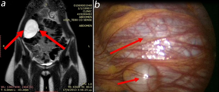
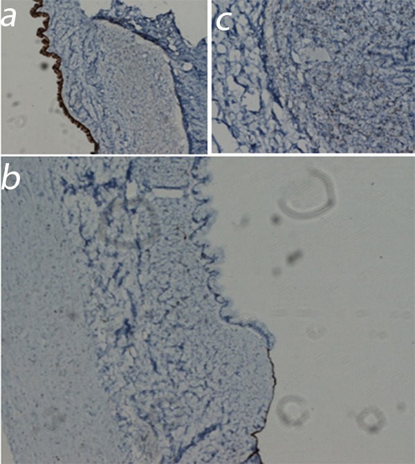
Case presentation: We present a 23-year-old woman, who was admitted to the hospital because of a palpable abdominal mass and discomfort since 4 months. Computed Tomography and Magnetic Resonance Imaging scans were performed and showed two retroperitoneal cystic masses, which were excised by laparoscopy. Histological and immunohistochemical examination revealed that the inner surfaces of the cysts were lined by epithelium with features of mesothelial cells, in addition to ovarian mucinous cystadenoma. This is the 29(th) case and the second reported case with two contemporary cysts.
Conclusion: The origin of retroperitoneal mucinous cystadenomas is still unclear. Pathological and immunohistochemical findings proved that these tumors resemble ovarian mucinous cystadenomas but are unattached to the ovary and can arise at any location in the retroperitoneum. Surgical excision of the aforementioned tumors is the treatment of choice. Hippokratia 2014; 18 (3): 278-281.
Keywords: Primary; cystadenoma; mucinous; retroperitoneal.
Figures
Similar articles
-
Primary Retroperitoneal Mucinous Cystadenoma.Ann Coloproctol. 2016 Feb;32(1):33-7. doi: 10.3393/ac.2016.32.1.33. Epub 2016 Feb 29. Ann Coloproctol. 2016. PMID: 26962534 Free PMC article.
-
Primary retroperitoneal mucinous cystadenoma-A case study and review of the literature.Int J Surg Case Rep. 2012;3(10):486-8. doi: 10.1016/j.ijscr.2012.05.010. Epub 2012 May 26. Int J Surg Case Rep. 2012. PMID: 22809878 Free PMC article.
-
Primary retroperitoneal mucinous cystadenoma.Saudi Med J. 2009 Jan;30(1):146-9. Saudi Med J. 2009. PMID: 19139790
-
A case report and a literature review of primary retroperitoneal mucinous cystadenoma: the importance of imaging in diagnosis and management.Future Oncol. 2018 Dec;14(28):2923-2931. doi: 10.2217/fon-2017-0649. Epub 2018 Jan 29. Future Oncol. 2018. PMID: 29376413 Review.
-
A unique benign mucinous cystadenoma of the retroperitoneum: a case report and review of the literature.Arch Gynecol Obstet. 2010 Jan;281(1):167-9. doi: 10.1007/s00404-009-1118-9. Epub 2009 May 16. Arch Gynecol Obstet. 2010. PMID: 19449022 Review.
Cited by
-
First Report of Retroperitoneal Mucinous Cystadenoma in a Patient with Hirsutism.Clin Med Res. 2020 Mar;18(1):27-32. doi: 10.3121/cmr.2019.1488. Epub 2019 Oct 3. Clin Med Res. 2020. PMID: 31582418 Free PMC article.
-
Retroperitoneal mucinous cystadenoma with neuroendocrine differentiation: a rare case and comprehensive approach to diagnosis and management.Diagn Pathol. 2025 May 3;20(1):58. doi: 10.1186/s13000-025-01658-7. Diagn Pathol. 2025. PMID: 40319316 Free PMC article.
-
Surgical resection of a rare primary retroperitoneal mucinous borderline tumor of Müllerian Origin: A case report.Gynecol Oncol Rep. 2022 Nov 8;44:101104. doi: 10.1016/j.gore.2022.101104. eCollection 2022 Dec. Gynecol Oncol Rep. 2022. PMID: 36388761 Free PMC article.
-
Primary Retroperitoneal Mucinous Tumours Diagnosed in Pregnancy: A Case Report and Literature Review.Int J Womens Health. 2019 Dec 20;11:649-653. doi: 10.2147/IJWH.S176219. eCollection 2019. Int J Womens Health. 2019. PMID: 31908544 Free PMC article.
-
Primary Retroperitoneal Mucinous Cystadenoma.Ann Coloproctol. 2016 Feb;32(1):33-7. doi: 10.3393/ac.2016.32.1.33. Epub 2016 Feb 29. Ann Coloproctol. 2016. PMID: 26962534 Free PMC article.
References
-
- Prabhuraj AR, Basu A, Sistla SC, Jaqdish S, Jayanthi S. Primary retroperitoneal mucinous cystadenoma in a man. Am J Clin Oncol. 2008;31:519–520. - PubMed
-
- Fujita N, Nishie A, Asayama Y, Kiyoshima K, Kubo Y, Honda H. A male case of primary retroperitoneal mucinous cystadenoma: a diagnostic dilemma. Jpn J Radiol. 2012;30:594–597. - PubMed
-
- Mattei J, Kim FJ, Phillips J, Zhelnin KE, Said S, Sehrt D, et al. Male primary retroperitoneal mucinous cystadenoma. Urology. 2013;82:e1–e2. - PubMed
-
- Lai KKT, Chan YYR, Chin ACW, Ng WF, Huang YHH, Mak YLM, et al. Primary retroperitoneal mucinous cystadenoma in a 52-year-old man. J HK Coll Radiol. 2004;7:223–225.
LinkOut - more resources
Full Text Sources