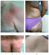
Mycosis fungoides in Iranian population: an epidemiological and clinicopathological study
- PMID: 25694829
- PMCID: PMC4324921
- DOI: 10.1155/2015/306543
Mycosis fungoides in Iranian population: an epidemiological and clinicopathological study
Abstract
Background. Mycosis fungoides (MF) is the most common subtype of cutaneous T-cell lymphoma. Extensive studies on Iranian MF patients are absent. The present study aimed to produce updated clinical information on Iranian MF patients. Methods. This was a retrospective, descriptive, single-center study, including all cases of MF seen in the Department of Dermatology, University Hospital of Isfahan, Iran, between 2003 and 2013. Data systematically recorded for each patient included clinical, biological, histological, and molecular findings. Results. Eighty-six patients with clinical and histologic diagnosis of MF were included in the study. Thirty-nine patients (45.3%) were male. Female predominance was observed in patients (male : female ratio is 1 : 1.2). Patients were between 7 and 84 years of age (median: 41). The interval from disease onset to diagnosis ranged from 0 to 55 years (median: 1 year). Eighteen cases (20.9%) had unusual variants of MF. The most common types included hypopigmented and poikilodermatous MF. Childhood cases of MF constituted 5.8% (5/86) of all patients. The early stages were seen in 82 cases (95.34%). Conclusion. The major differences in epidemiologic characteristics of MF in Iran are the lack of male predominance and the lower age of patients at the time of diagnosis.
Figures

References
-
- Siegel R. S., Pandolfino T., Guitart J., Rosen S., Kuzel T. M. Primary cutaneous T-cell lymphoma: review and current concepts. Journal of Clinical Oncology. 2000;18(15):2908–2925. - PubMed
LinkOut - more resources
Full Text Sources
Other Literature Sources

