

Heart Rate Variability: New Perspectives on Physiological Mechanisms, Assessment of Self-regulatory Capacity, and Health risk
- PMID: 25694852
- PMCID: PMC4311559
- DOI: 10.7453/gahmj.2014.073
Heart Rate Variability: New Perspectives on Physiological Mechanisms, Assessment of Self-regulatory Capacity, and Health risk
Abstract
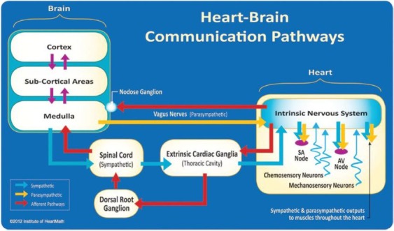
Heart rate variability, the change in the time intervals between adjacent heartbeats, is an emergent property of interdependent regulatory systems that operates on different time scales to adapt to environmental and psychological challenges. This article briefly reviews neural regulation of the heart and offers some new perspectives on mechanisms underlying the very low frequency rhythm of heart rate variability. Interpretation of heart rate variability rhythms in the context of health risk and physiological and psychological self-regulatory capacity assessment is discussed. The cardiovascular regulatory centers in the spinal cord and medulla integrate inputs from higher brain centers with afferent cardiovascular system inputs to adjust heart rate and blood pressure via sympathetic and parasympathetic efferent pathways. We also discuss the intrinsic cardiac nervous system and the heart-brain connection pathways, through which afferent information can influence activity in the subcortical, frontocortical, and motor cortex areas. In addition, the use of real-time HRV feedback to increase self-regulatory capacity is reviewed. We conclude that the heart's rhythms are characterized by both complexity and stability over longer time scales that reflect both physiological and psychological functional status of these internal self-regulatory systems.
心率变异性 (HRC)(在相邻心跳之 间的时间间隔的变化)是相互依存 的调节系统的一种紧急特性,在不 同时段内发生,以适应环境和心理 挑战。这篇文章简要回顾了心脏的 神经调节,在心率变异性的极低频 率节奏机制方面,提供了一些新的 观点。在健康风险和生理及心理自 我调节能力评估讨论的背景下解读 HRV 节律。伴有心血管系统输入功 能的更高级的大脑中枢集成输入的 脊髓和延髓心血管监管中心,通过 交感和副交感神经传出通路调节心 率和血压。我们还讨论了先天性心 脏神经系统和心脏大脑连接通路, 借以传入信息并可以影响皮质下 区,皮质前区和运动皮质区的活 动。此外,审查了使用实时的 HRV 反馈以提高自我调节能力。我们的 结论是,心脏节律的特征在于在更 长的时段上既有复杂性又有稳定 性,这反映出这些内部自我调节系 统的生理和心理功能状态。
La variabilidad de la frecuencia cardiaca, o modificación de los intervalos de tiempo entre los latidos consecutivos del corazón, es una propiedad emergente de los sistemas reguladores interdependientes que opera sobre diferentes escalas temporales para adaptarse a los retos ambientales y psicológicos. Este artículo revisa brevemente la regulación nerviosa del corazón y ofrece nuevas perspectivas sobre los mecanismos subyacentes al ritmo de muy baja frecuencia de la variabilidad de la frecuencia cardiaca. Se analiza la interpretación de los ritmos de la variabilidad de la frecuencia cardiaca en el contexto del riesgo para la salud y la valoración de la capacidad autorregulatoria fisiológica y psicológica. Los centros reguladores cardiovasculares de la médula espinal y del bulbo raquídeo integran entradas de centros cerebrales superiores con entradas de sistemas cardiovasculares aferentes para ajustar la frecuencia cardiaca y la tensión arterial por vías eferentes simpáticas y parasimpáticas. También hablamos sobre el sistema cardiaco nervioso intrínseco y las vías de conexión corazón-cerebro, a través de las cuales la información aferente puede influir sobre la actividad en las áreas subcortical, frontocortical y de la corteza motora. Además, se revisa el uso de retroalimentación de variabilidad de la frecuencia cardiaca a tiempo real para aumentar la capacidad autorreguladora. Concluimos que los ritmos cardiacos se caracterizan tanto por su complejidad como por su estabilidad sobre escalas temporales más largas que reflejan los estados funcionales tanto fisiológicos como psicológicos de estos sistemas internos autorreguladores.
Keywords: Heart rate variability; health risk; physiological mechanisms; self-regulatory capacity.
Figures
References
-
- Cannon WB. The James-Lange theory of emotion: a critical examination and an alternative theory. Am J Psychol. 1987;100(3–4):567–86. - PubMed
-
- Singer DH, Martin GJ, Magid N, et al. Low heart rate variability and sudden cardiac death. J Electrocardiol. 1988; 21 Suppl: S46–55. - PubMed
-
- McCraty R, Atkinson M, Tomasino D, Bradley RT. The coherent heart: heart-brain interactions, psychophysiological coherence, and the emergence of system-wide order. Boulder Creek, CA: Institute of Heartmath; 2009.
-
- McCraty R, Childre D. Coherence: bridging personal, social, and global health. Altern Ther Health Med. 2010;16(4):10–24. - PubMed
Publication types
LinkOut - more resources
Full Text Sources
Other Literature Sources
Medical
