. 2015:2015:206382.
doi: 10.1155/2015/206382. Epub 2015 Jan 28.

A complex genome-microRNA interplay in human mitochondria

Affiliations

A complex genome-microRNA interplay in human mitochondria

Santosh Shinde et al. Biomed Res Int. 2015.

Abstract

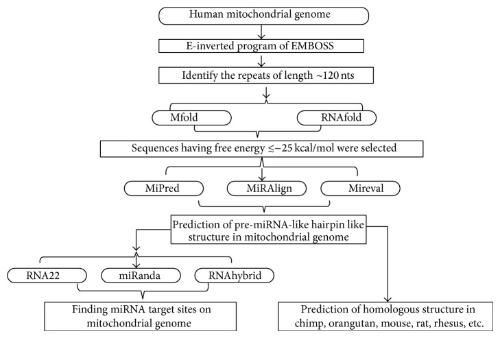
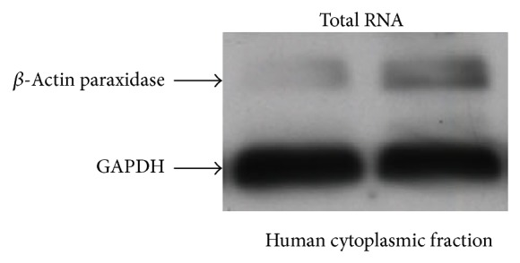
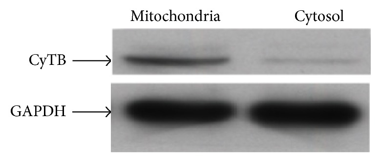
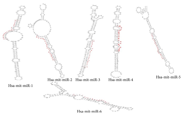
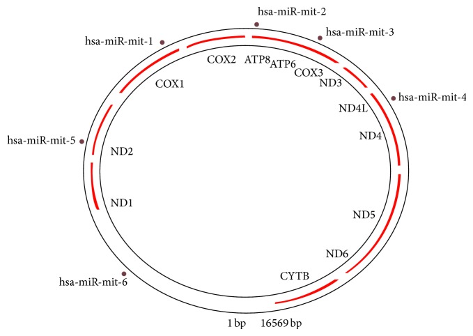
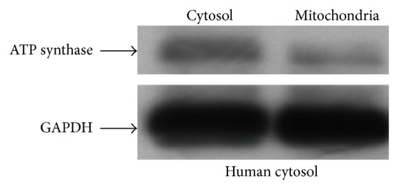
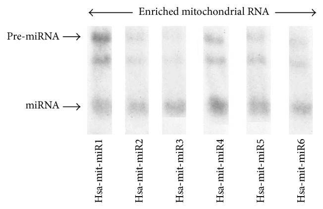
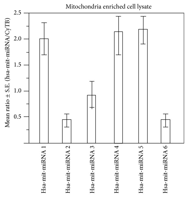
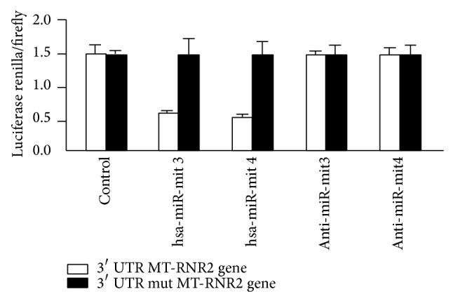
Small noncoding regulatory RNA exist in wide spectrum of organisms ranging from prokaryote bacteria to humans. In human, a systematic search for noncoding RNA is mainly limited to the nuclear and cytosolic compartments. To investigate whether endogenous small regulatory RNA are present in cell organelles, human mitochondrial genome was also explored for prediction of precursor microRNA (pre-miRNA) and mature miRNA (miRNA) sequences. Six novel miRNA were predicted from the organelle genome by bioinformatics analysis. The structures are conserved in other five mammals including chimp, orangutan, mouse, rat, and rhesus genome. Experimentally, six human miRNA are well accumulated or deposited in human mitochondria. Three of them are expressed less prominently in Northern analysis. To ascertain their presence in human skeletal muscles, total RNA was extracted from enriched mitochondria by an immunomagnetic method. The expression of six novel pre-miRNA and miRNA was confirmed by Northern blot analysis; however, low level of remaining miRNA was found by sensitive Northern analysis. Their presence is further confirmed by real time RT-PCR. The six miRNA find their multiple targets throughout the human genome in three different types of software. The luciferase assay was used to confirm that MT-RNR2 gene was the potential target of hsa-miR-mit3 and hsa-miR-mit4.


Figures

Figure 1
Figure 1
Flow chart of miRNA prediction in mitochondrial genome (bioinformatics analysis).
Figure 2
Figure 2
Blot showing quantitative Western blot analysis of β-actin peroxidase in relation to loading differences of GAPDH probes extracted from whole cell total RNA.
Figure 3
Figure 3
Blots showing mitochondrial probe CyTB in enriched mitochondrial and cytosolic RNA loading in two different lanes of the gel. The CyTB differential expression is compared to GAPDH loading.
Figure 4
Figure 4
Predicted pre-miRNA and miRNA structure.
Figure 5
Figure 5
Schematic presentation of human miRNA prediction in mitochondrial genome.
Figure 6
Figure 6
Western blot hybridization with ATP synthase isolated from mitochondria enriched and cytosolic RNA.
Figure 7
Figure 7
Autoradiogram showing Northern blots hybridization probed with six different pre-miRNA antisense probes. Northern blot was performed with equal amount of enriched mitochondrial RNA.
Figure 8
Figure 8
Ratio of the six miRNA in comparison to CyTB. Relative ratios in triplicate were calculated following RT-PCR. The height of the bar is proportional to the amount of human mitochondrial miRNA.
Figure 9
Figure 9
Intersection of miRanda, PicTar, and TargetScan results.
Figure 10
Figure 10
Schematic representation of common miRNA target sites on mitochondrial genome.
Figure 11
Figure 11
Schematic representation of miRNA conserved target on mitochondria (miRanda versus RNA22).
Figure 12
Figure 12
Interaction map of miRNA and mitochondrial genome. Interactions among mitochondrial genome and miRNA are depicted with arrows, number on that arrow represents number of miRNA targets predicted by the software, and different colors of arrow represent type of software.
Figure 13
Figure 13
Luciferase assay of hsa-miR-mit3 and has-miR-mit4. Luciferase assay was used to invent whether MT-RNR2 was the direct target of hsa-miR-mit3 or hsa-miR-mit4 microRNA separately. A wild type and a mutated 3′ UTR of MT-RNR2 gene were subcloned into the psiCHECK-2 luciferase miRNA expression reporter vector as described earlier [23]. The transfected cells were cotransfected with 100 mM anti-hsa-miR-mit3 or the same amount of anti-hsa-miR-mit4.
Figure 14
Figure 14
miRNA detected in three increasing mitochondrial mt-RNA inputs.

References

    1. Anderson S., Bankier A. T., Barrell B. G., et al. Sequence and organization of the human mitochondrial genome. Nature. 1981;290(5806):457–465. doi: 10.1038/290457a0. - DOI - PubMed
    1. Ballinger S. W., Shoffner J. M., Hedaya E. V., et al. Maternally transmitted diabetes and deafness associated with a 10.4 kb mitochondria DNA deletion. Nature Genetics. 1992;1(1):11–15. doi: 10.1038/ng0492-11. - DOI - PubMed
    1. Brown M. D., Wallace D. C. Spectrum of mitochondrial DNA mutations in Leber's hereditary optic neuropathy. Clinical Neuroscience. 1994:138–145.
    1. Easteal S. The relative rate of DNA evolution in primates. Molecular Biology and Evolution. 1991;8(1):115–127. - PubMed
    1. Johns D. R., Neufeld M. J. Cytochrome C oxidase mutations in Leber hereditary optic neuropathy. Biochemical and Biophysical Research Communications. 1993;196(2):810–815. doi: 10.1006/bbrc.1993.2321. - DOI - PubMed

Publication types

LinkOut - more resources