. 2015 Mar 12;58(5):2378-89.
doi: 10.1021/jm501859j. Epub 2015 Feb 26.

Development of a novel class of tubulin inhibitor from desmosdumotin B with a hydroxylated bicyclic B-ring

Affiliations

Development of a novel class of tubulin inhibitor from desmosdumotin B with a hydroxylated bicyclic B-ring

Kyoko Nakagawa-Goto et al. J Med Chem. .

Abstract

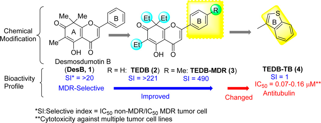
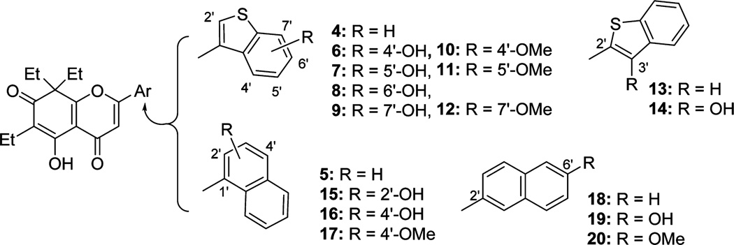
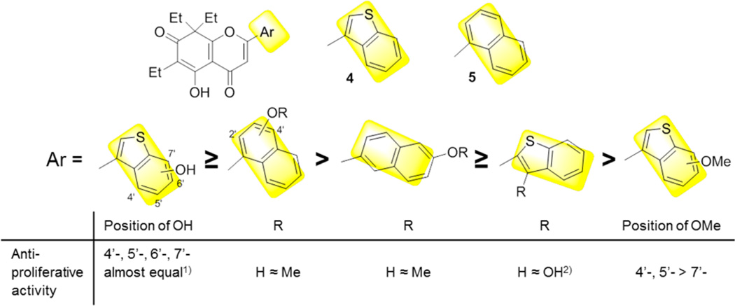
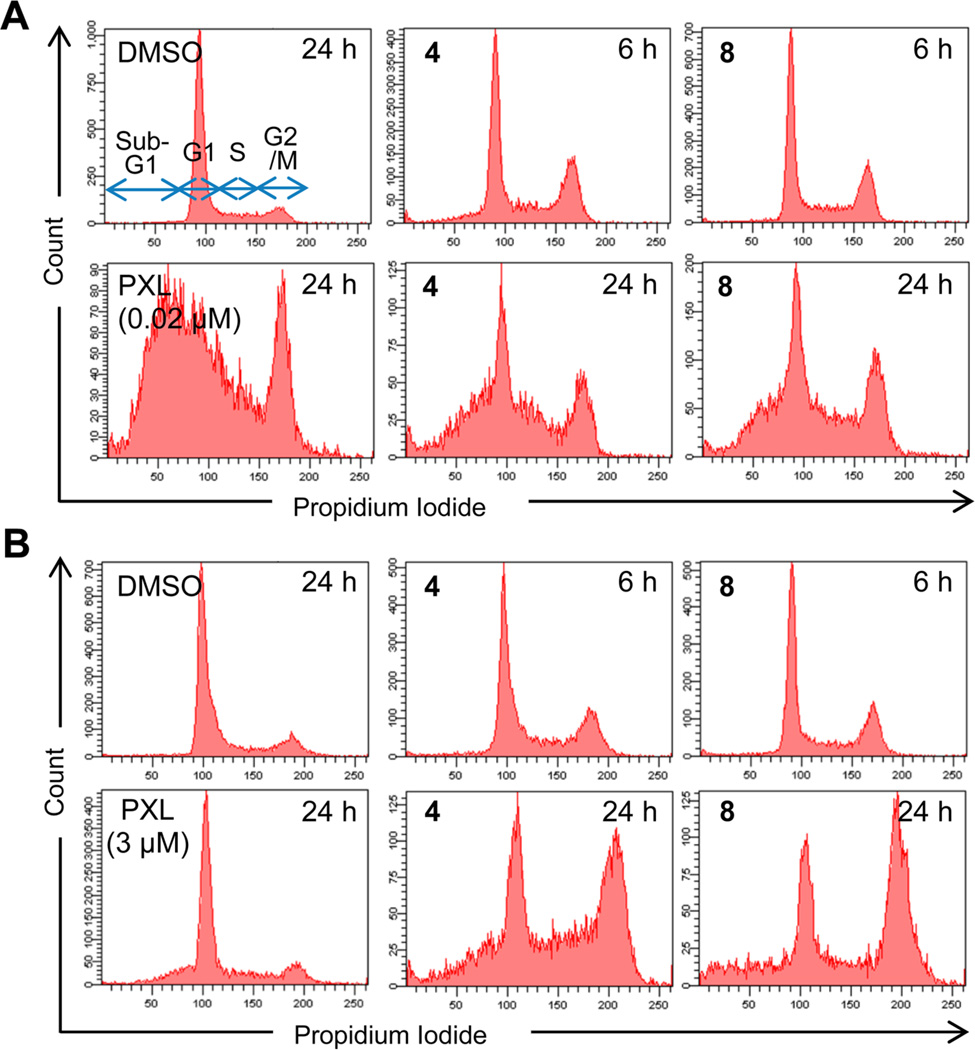
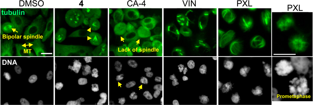
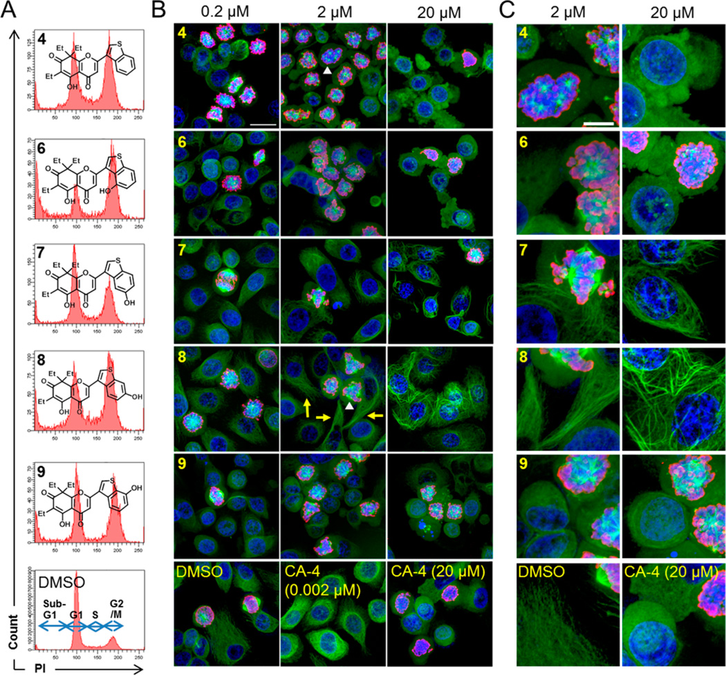
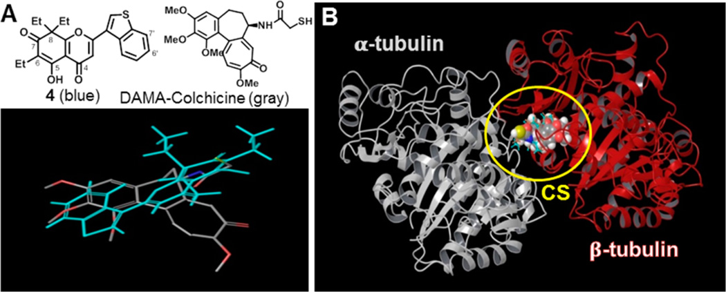
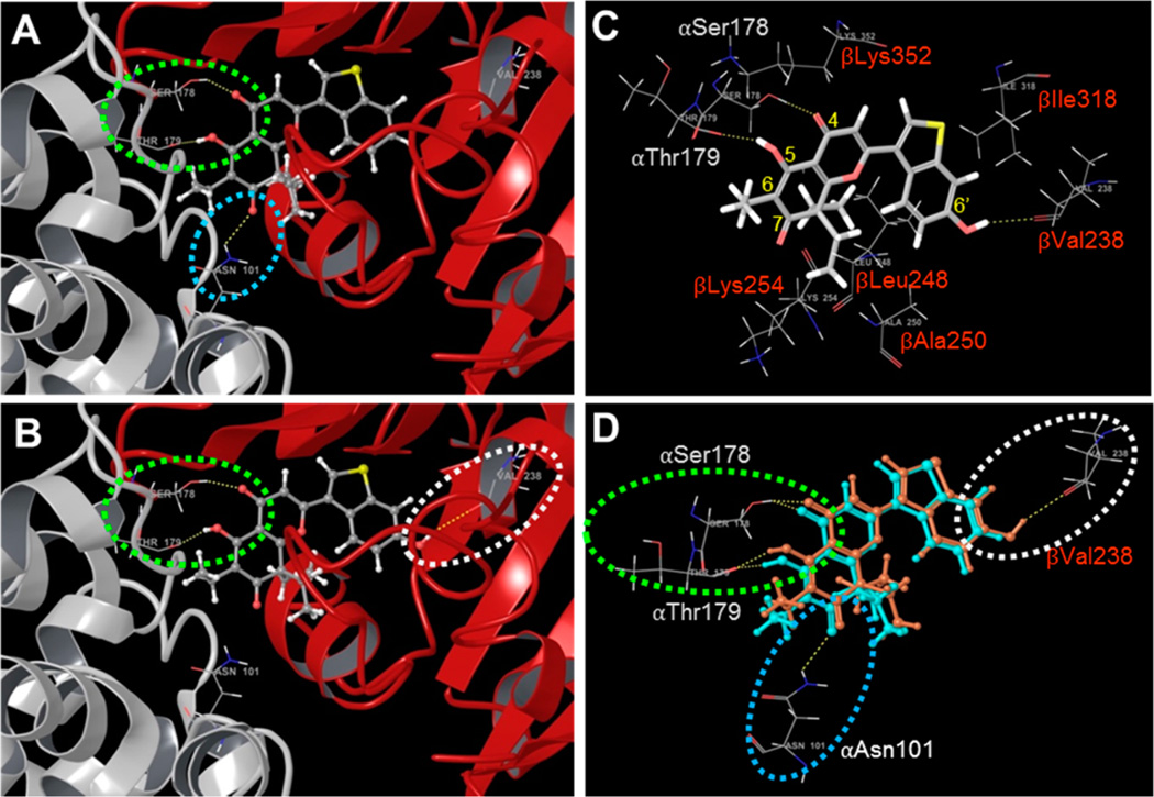
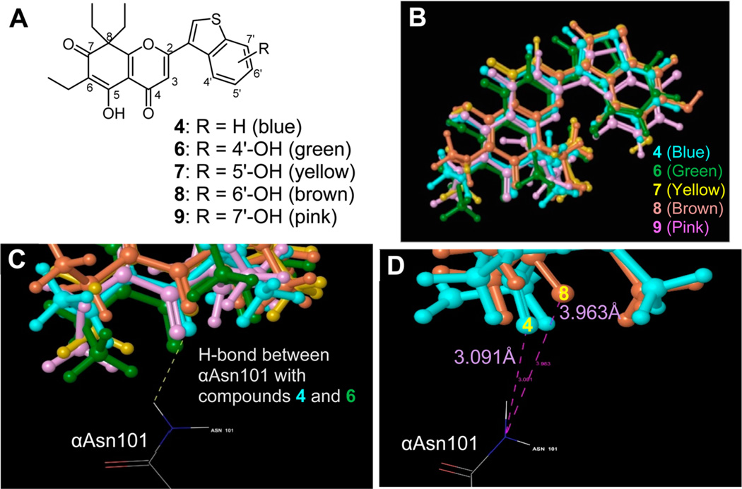
A series of newly synthesized hydroxylated analogues of triethyldesmosdumotin B (TEDB) with a bicyclic B-ring exhibited a significantly different mode of action for affecting microtubule dynamics and spindle formation but had the same antiproliferative activity spectrum, including activity against multidrug-resistant tumors. These analogues efficiently induced cell cycle arrest at prometaphase and caused formation of immature multipolar spindles. 6'-Hydroxyl TEDB-TB (8) disrupted bipolar spindle formation but had a negligible effect on interphase microtubules. On the basis of the predicted binding modes of the new compounds with tubulin dimer, compound 4 forms three hydrogen bonds (H-bonds) only with α-tubulin at the colchicine site; in contrast, 8 forms H-bonds with both α- and β-tubulin. We predict that, when a compound/ligand, such as 8, forms H-bonds to both α- and β-tubulins, spindle formation is disrupted more than the dynamics of interphase microtubules. This result may reflect the well-known greater dynamicity of spindle microtubules as compared with interphase microtubules.


Figures

Figure 1
Figure 1
Structures of desmosdumotin B and related bioactive analogues.
Figure 2
Figure 2
Structures of TEDB-TB analogues.
Figure 3
Figure 3
Structure-activity relationship of hydroxylated TEDB-TB analogues. SAR study shown in Table 1 summarized: (1) 4′-position ≈ 6′-position > 5′-position ≈ 7′-position against HCT-8 and (2) H > OH against PC-3.
Figure 4
Figure 4
Effect of compounds on the cell cycle distribution in MDR cells. Chemosensitive KB cell line (A) and its MDR subline (KB-VIN) (B) were treated with vehicle control (DMSO), PXL (0.02 µM for KB and 3 µM for KB-VIN), 4, or 8 for 6 or 24 h, as indicated. Cell cycle progression was analyzed by measuring DNA content using a flow cytometer. Cell cycle phases are indicated as G1 (2N), S, G2/M (4N), and sub-G1. PXL disrupted cell cycle progression in KB cells at 0.02 µM, while no significant effect was seen in KB-VIN cells, even at 3 µM. Accumulation of G2/M phase as well as S phase cells was observed in both chemosensitive and MDR cells treated with 4 and 8 at the same concentrations in a time-dependent manner.
Figure 5
Figure 5
Disruption of microtubule morphology in PC-3 cells by small molecules. Prostate cancer cells (PC-3) were treated for 24 h with antimicrotubule agents as indicated (DMSO as vehicle control, 0.2 µM compound 4, 0.2 µM CA-4, 5 µM VIN, 0.2 µM PXL). Compounds were used at equitoxic compound concentrations and examined for disruption of microtubule dynamics within 24 h. Microtubules were visualized by immunofluorescence staining using monoclonal antibody to α-tubulin (upper panels), and DAPI was used for DNA staining (lower panels). In control cells treated with DMSO, bipolar spindle formation in mitotic cells (arrow) as well as microtubule (MT) networks in interphase cells (double arrow) were observed. Treatment with 4 induced depolymerization of cytosolic microtubules as well monopolar (arrow heads) or multipolar spindle formations in the G2/M-phase. Treatment with CA-4 led to both depolymerization of interphase microtubules and disruption of mitotic spindles (arrows). PXL inhibited the onset of mitosis and arrested cells at prometaphase with condensed chromosomes (right panels). Characteristic bundled microtubules were observed in interphase cells treated with PXL, while tubulin paracrystal formation was observed in cells treated with VIN. Bar, 25 µm. Additional images including colchicine treatment are shown in the Supporting Information (Figure S1).
Figure 6
Figure 6
Distinct effects on microtubule morphology by structural modification of compounds. (A) Effect of compounds on cell cycle distribution in PC-3 cells. PC-3 cells were treated for 24 h with 2 µM 4, 6, 7, 8, or 9 or vehicle control (DMSO), as indicated. Cell cycle progression was analyzed by flow cytometric analysis. All analogues efficiently induced accumulation of G2/M phase cells. (B) Dose-dependent effects of compounds on microtubule morphology in PC-3 cells. PC-3 cells were treated for 24 h with 4, 6, 7, 8, or 9 at 0.2, 2, or 20 µM. DMSO was used with the control cells. CA-4 was used at 0.002 or 20 µM. Cells were stained with antibodies to α-tubulin (green) and Ser10-phosphorylated histone H3 (p-H3, red) as a mitotic marker and with DAPI for DNA (blue). Stacked and merged confocal images are presented. TEDB-TB analogues induced cell cycle arrest at prometaphase (p-H3-positive) by disrupting bipolar spindle formation. Immature multipolar spindles (white arrow heads) were formed in the cells treated with analogues but were undetectable after CA-4 treatment. Dose-dependent disruption of both interphase microtubules and spindles was observed in cells treated with 4, 6, 9, or CA-4. In contrast, in cells treated with 7 or 8, normal interphase microtubules and disrupted spindles were visualized (yellow arrows). Bar, 25 µm. (C) Higher magnification views. Bar, 10 µm. Additional images are shown in the Supporting Information (Figure S2).
Figure 7
Figure 7
Predicted docking models for 4 and 8 binding to tubulin. (A) Structures and 3D models of 4 (blue skeleton) and DAMA-colchicine (gray skeleton with oxygen in red, nitrogen in blue, and sulfur in yellow). (B) Docking model of 4 (blue skeleton), 8 (brown skeleton), and DAMA-colchicine (sphere in 3D with carbon in gray, proton in white, oxygen in red, nitrogen in blue, and sulfur in yellow) in the CS (yellow circle) of the tubulin crystal structure (PDB ID: 1SA0), shown as a ribbon diagram.
Figure 8
Figure 8
Predicted docking models for 4 and 8 binding in the CS. Crystal structures (PDB ID: 1SA0) of α- (white) and β-tubulin (red) are shown as ribbon diagrams. H-bonds calculated to be less than 3 Å between protein and compounds are represented by dashed lines. Docking models of compounds (gray skeleton with oxygen in red and sulfur in yellow) 4 (A) and 8 (B) in the CS are shown. Superimposition of docked compound 4 shows H-bonds with the side chains of αAsn101 (dashed blue circle) and αSer178 and αThr179 (dashed green circle). In contrast, with 8, there is a H-bond between 8 and the side chain of βVal238 (dashed white circle), and the H-bond between 8 and the side chain of αAsn101 is absent. (C) Docking mode of 8 in the CS. Superimposition of docked compound 8 shows H-bonds between 8 and αSer178 and αThr179 in α-tubulin (αSer178, αThr179) and between 8 and βVal238. (D) Comparison of the docking mode of 8 (brown) with that of 4 (blue) in CS. Superimposition of docked compounds 4 and 8 shows conserved H-bonds with αSer178 and αThr179 (dashed green circle). An additional H-bond between the C-7 oxygen of 4 and αAsn101 (dashed blue circle) was also observed. In contrast, a H-bond between the C-6′ hydroxyl group of 8 and the side chain of βVal238 (white circle) was unambiguous, while no H-bond occurred with αAsn101.
Figure 9
Figure 9
Hydrogen bonds between 4 analogues and tubulin in the CS. Structures (A) and conformations (B) of 4 and analogues are presented. The same docking pattern is shown among all compounds, except 9. (C) H-bond between C-7 oxygen of compounds and αAsn101. A H-bond calculated as less than 3 Å occurred in 4 and 6. (D) Comparison of distance between oxygen on compound and nitrogen on αAsn101. The N-O distances for 4 (3.091 Å) and 8 (3.963 Å) differed by over 0.8 Å.
Scheme 1
Scheme 1
Syntheses of New Analogues of 4 and 5a aReagents and conditions: (a) ArCHO, 50% KOH, EtOH, rt; (b) I2 (cat.), DMSO, H2SO4 (cat.), 90–95 °C, 1 h; (c) BBr3, 0 °C to rt.
Scheme 2
Scheme 2
Preparation of Methoxybenzothiophenecarboxaldehydesa aReagents and conditions: (a) CH3COCH2Cl, K2CO3, DMF, rt; (b) PPA, 100 °C; (c) NBS, (PhCOO)2, CCl4, reflux; (d) HMTA, CHCl3, reflux, then 50% HOAc, reflux; (e) NaOMe, CuO, CuI, KI, DMF, reflux; (f) n-BuLi, THF, −78 °C, then DMF.

References

    1. Dumontet C, Jordan MA. Microtubule-binding agents: A dynamic field of cancer therapeutics. Nat. Rev. Drug Discovery. 2010;9:790–803. - PMC - PubMed
    1. Risinger AL, Giles FJ, Mooberry SL. Microtubule dynamics as a target in oncology. Cancer Treat. Rev. 2009;35:255–261. - PMC - PubMed
    1. Jordan MA, Wilson L. Microtubules as a target for anticancer drugs. Nat. Rev. Cancer. 2004;4:253–265. - PubMed
    1. Lu Y, Chen J, Xiao M, Li W, Miller DD. An overview of tubulin inhibitors that interact with the colchicine binding site. Pharm. Res. 2012;29:2943–2971. - PMC - PubMed
    1. Eckford PD, Sharom FJ. ABC efflux pump-based resistance to chemotherapy drugs. Chem. Rev. 2009;109:2989–3011. - PubMed

Publication types

MeSH terms

LinkOut - more resources