. 2015 May 1;467(3):453-60.
doi: 10.1042/BJ20141248.

Regulation of IGFBP-2 expression during fasting

Affiliations

Regulation of IGFBP-2 expression during fasting

Hye Suk Kang et al. Biochem J. .

Abstract

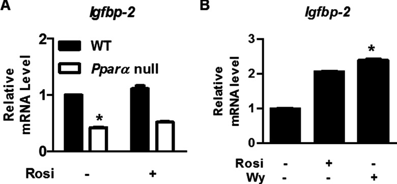
Insulin-like growth factor (IGF)-binding protein-2 (IGFBP-2), one of the most abundant circulating IGFBPs, is known to attenuate the biological action of IGF-1. Although the effect of IGFBP-2 in preventing metabolic disorders is well known, its regulatory mechanism remains unclear. In the present study, we demonstrated the transcriptional regulation of the Igfbp-2 gene by peroxisome-proliferator-activated receptor (PPAR) α in the liver. During fasting, both Igfbp-2 and PPARα expression levels were increased. Wy14643, a selective PPARα agonist, significantly induced Igfbp-2 gene expression in primary cultured hepatocytes. However, Igfbp-2 gene expression in Pparα null mice was not affected by fasting or Wy14643. In addition, through transient transfection and chromatin immunoprecipitation assay in fasted livers, we determined that PPARα bound to the putative PPAR-responsive element between -511 bp and -499 bp on the Igfbp-2 gene promoter, indicating that the Igfbp-2 gene transcription is activated directly by PPARα. To explore the role of PPARα in IGF-1 signalling, we treated primary cultured hepatocytes with Wy14643 and observed a decrease in the number of IGF-1 receptors (IGF-1Rs) and in Akt phosphorylation. No inhibition was observed in the hepatocytes isolated from Pparα null mice. These results suggest that PPARα controls IGF-1 signalling through the up-regulation of hepatic Igfbp-2 transcription during fasting and Wy14643 treatment.


Figures

Figure 1
Figure 1. Microarray analysis to identify Igfbp-2 gene expression level and qPCR analysis in fasting condition
(A) Hierarchical gene clustering was generated with the TM4 Microarray Software Suite (MeV) from fasted and refed livers. A heat map showing significant changes in a group of selected genes from the livers of mice that were fasted for 24 h and refed for 12 h. The ratios of gene profiles are presented as a heat map (left panel) and gene expression pattern (right panel). Fold change and P-values of the genes in the clusters are given on the right. (BE) mRNA levels of Igfbp-1, Igfbp-2, Pparα and Pck1 in the liver of WT mice were measured by qPCR under the indicated conditions. **P<0.01 and ***P<0.001 compared with fed mice.
Figure 2
Figure 2. PPARα is involved in the induction of IGFBP-2 following the fasting state and Wy14643 treatment
(A) Mouse primary hepatocytes were treated with Wy14643 (Wy) for 6 h at the indicated concentrations. Total RNA was isolated and analysed using qPCR with the observed primers. (B) Mouse primary hepatocytes from WT and Pparα null mice were treated with or without Wy14643 for 6 h. The level of Pparα mRNA was measured by qPCR analysis. (C) The mRNA level of Igfbp-2 in liver from WT and Pparα null mice fed and fasted for 24 h was analysed by qPCR. (D) Protein level of IGFBP-2 in livers from WT and Pparα null mice fed and fasted for 24 h was analysed by Western blot analysis. (E) Secretion level of IGFBP-2 in vivo and in vitro. Mice were fed or fasted for 24 h and serum was collected from WT and Pparα null mice. Secretion levels of IGFBP-2 in feeding or fasting conditions were measured by ELISA. (F) WT and Pparα null mice were treated with or without Wy14643 for 6 h. Whole cell extracts were isolated from primary hepatocytes of the observed conditions and assessed by Western blot analysis with the indicated antibody. (GI) Primary hepatocytes from WT and Pparα null mice were treated with or without Wy14643 for 6 h. The level of Pparγ1, PPARγ2 and PPARδ mRNA was measured by qPCR analysis. *P<0.05 and **P<0.01 compared with untreated control or fed WT mice.
Figure 3
Figure 3. Identification of PPRE in the mIgfbp-2 promoter
(A) Comparison of putative PPRE with consensus PPRE sequences. A proposed PPARα-binding element is shaded on the Igfbp-2 promoter between −511 and −499. The numbers indicate the distance in nucleotides from the transcription start site (+1) of the mouse Igfbp-2 gene. (B) Effects of PPARα on promoter reporter activities in deletion constructs of the Igfbp-2 gene. Deletion construct of the mIgfbp-2 promoter was transiently co-transfected with pcDNA3 (open bars) or PPARα expression vector (closed bars) in HEK-293T cells. After 24 h, media were changed to contain 20 μM Wy14643. Luciferase activity was normalized to β-galactosidase activity to correct for transfection efficiency. (C) Internal deletion constructs for the Igfbp-2 promoter were analysed for promoter activity. (D and E) ChIP assay. Mice were fasted for 24 h and refed for 12 h. Chromatins were isolated from mice livers and ChIP assay was performed. Input represents 10% of purified DNA in each sample. Nuclear extracts from mice livers were immunoprecipitated with anti-PPARα antibody and purified DNA samples were used to perform qPCR with primers binding to the putative PPRE regions on the mIgfbp-2 (D) and mG6pc (E) gene promoters. All data are representative of at least three independent experiments. *P<0.05 compared with untreated control.
Figure 4
Figure 4. The IGF-1 signalling system is mediated by PPARα in primary hepatocytes
WT and Pparα null mice were pretreated with Wy14643 for 6 h and then exposed to IGF-1 for 15 min under the indicated conditions. Whole cell extracts were isolated from primary hepatocytes of the indicated groups and assessed by Western blot analysis with various antibodies. Right panel indicate the density of Western blot bands measured with ImageJ software. *P<0.05 compared with untreated control or IGF-1-treated cells and #P<0.05 compared with IGF-1- and Wy14643-treated cells.
Figure 5
Figure 5. Effects of PPAR agonists in the primary hepatocytes and hepatic cell lines
(A) Effect of rosiglitazone on Igfbp-2 gene expression. Rosiglitazone (10 μM) was added to primary hepatocytes for 6 h and RNA was harvested for qPCR. (B) mRNA levels of Igfbp-2 after rosiglitazone or Wy14643 treatment in the HepG2 cell line. Rosiglitazone (10 μM) or Wy14643 (10 μM) was added to HepG2 cells for 6 h and RNA was harvested for qPCR. Values are expressed as mean values±S.E.M. from three experiments. Rosi, rosiglitazone; Wy, Wy14643. *P<0.05 compared with untreated group.

References

    1. Hwa V., Oh Y., Rosenfeld R.G. The insulin-like growth factor-binding protein (IGFBP) superfamily. Endocr. Rev. 1999;20:761–787. - PubMed
    1. Jones J.I., Clemmons D.R. Insulin-like growth factors and their binding proteins: biological actions. Endocr. Rev. 1995;16:3–34. - PubMed
    1. Oh Y. IGF-independent regulation of breast cancer growth by IGF binding proteins. Breast Cancer Res. Treat. 1998;47:283–293. doi: 10.1023/A:1005911319432. - DOI - PubMed
    1. Ricort J.M. Insulin-like growth factor binding protein (IGFBP) signalling. Growth Horm. IGF Res. 2004;14:277–286. doi: 10.1016/j.ghir.2004.02.002. - DOI - PubMed
    1. Wheatcroft S.B., Kearney M.T. IGF-dependent and IGF-independent actions of IGF-binding protein-1 and -2: implications for metabolic homeostasis. Trends Endocrinol. Metab. 2009;20:153–162. doi: 10.1016/j.tem.2009.01.002. - DOI - PubMed

Publication types

MeSH terms