. 2015 May 1;113(9):3268-80.
doi: 10.1152/jn.00970.2014. Epub 2015 Feb 18.

Repetitive mild traumatic brain injury induces ventriculomegaly and cortical thinning in juvenile rats

Affiliations

Repetitive mild traumatic brain injury induces ventriculomegaly and cortical thinning in juvenile rats

Corey Goddeyne et al. J Neurophysiol. .

Abstract

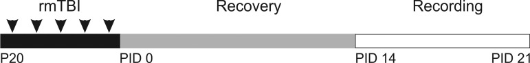
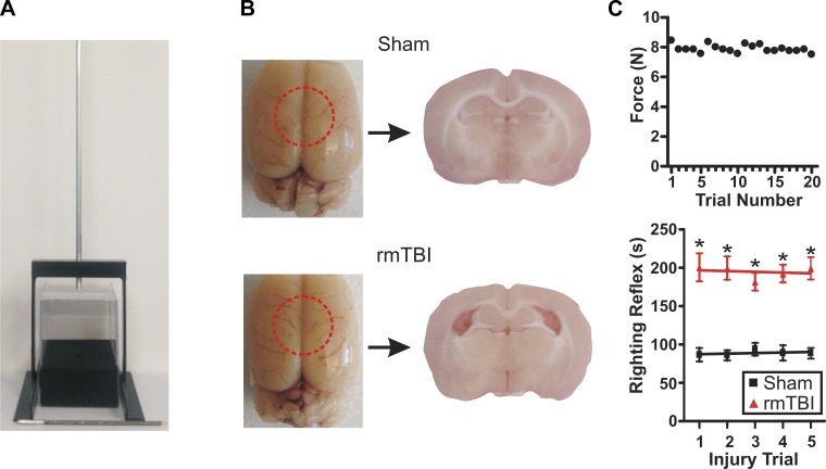
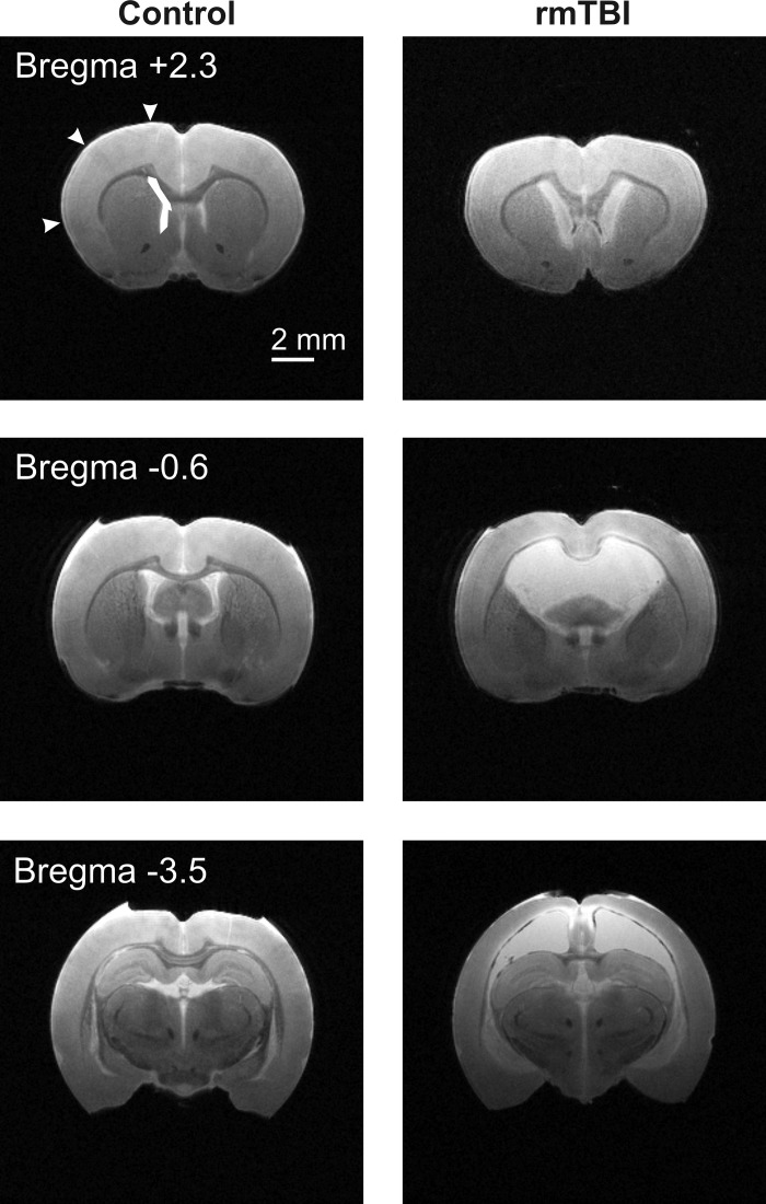
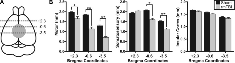
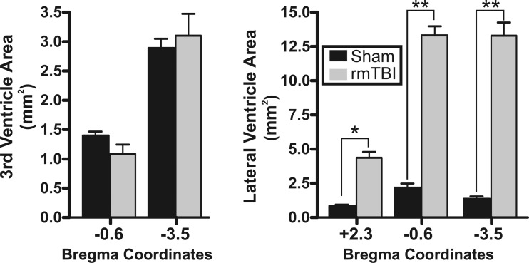
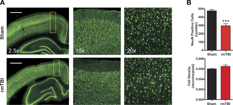
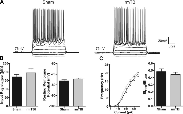
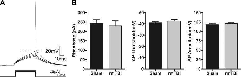
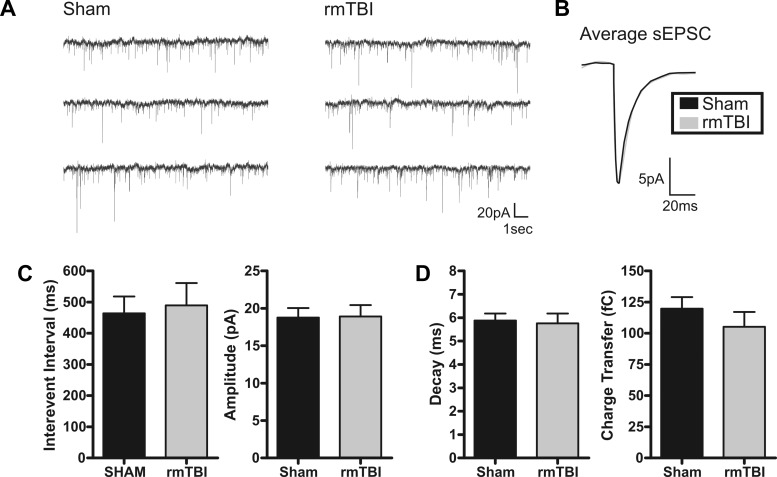
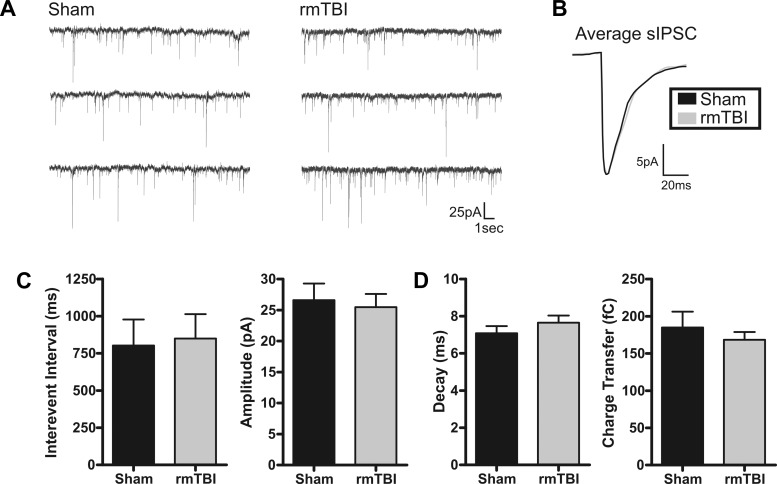
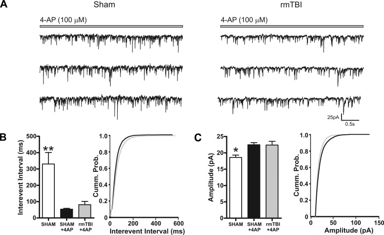
Traumatic brain injury (TBI) most frequently occurs in pediatric patients and remains a leading cause of childhood death and disability. Mild TBI (mTBI) accounts for nearly 75% of all TBI cases, yet its neuropathophysiology is still poorly understood. While even a single mTBI injury can lead to persistent deficits, repeat injuries increase the severity and duration of both acute symptoms and long-term deficits. In this study, to model pediatric repetitive mTBI (rmTBI) we subjected unrestrained juvenile animals (postnatal day 20) to repeat weight-drop impacts. Animals were anesthetized and subjected to sham injury or rmTBI once per day for 5 days. Magnetic resonance imaging (MRI) performed 14 days after injury revealed marked cortical atrophy and ventriculomegaly in rmTBI animals. Specifically, beneath the impact zone the thickness of the cortex was reduced by up to 46% and the area of the ventricles increased by up to 970%. Immunostaining with the neuron-specific marker NeuN revealed an overall loss of neurons within the motor cortex but no change in neuronal density. Examination of intrinsic and synaptic properties of layer II/III pyramidal neurons revealed no significant difference between sham-injured and rmTBI animals at rest or under convulsant challenge with the potassium channel blocker 4-aminopyridine. Overall, our findings indicate that the neuropathological changes reported after pediatric rmTBI can be effectively modeled by repeat weight drop in juvenile animals. Developing a better understanding of how rmTBI alters the pediatric brain may help improve patient care and direct "return to game" decision making in adolescents.

Keywords: MRI; concussion; electrophysiology; pediatrics; traumatic brain injury.


Figures

Fig. 1.
Fig. 1.
Experimental timeline. Overview of the timeline used to model repetitive mild traumatic brain injury (rmTBI). Arrowheads represent time of single impact repeated once daily for 5 days. Control animals were given anesthesia only. Postinjury day (PID) indicates number of days after the 5th rmTBI injury.
Fig. 2.
Fig. 2.
Experimental model of rmTBI. A: photograph of rmTBI device and impact weight. B, left: photographs of brains acutely prepared 14 days after sham injury or rmTBI (1 impact/day for 5 days) in juvenile (P20) rats. Red dashed circle indicates approximate site of impact. Right: photograph of coronal brain slices taken from respective sham-injured or rmTBI brains. Note the presence of enlarged ventricles and cortical thinning after rmTBI. C, top: scatterplot of impact force measurements taken across 20 trials (weight 92 g, drop height 865 mm). Bottom: line graph of average righting reflex time in sham-injured (n = 38) and rmTBI (n = 42) animals across the 5 injury trial days. *P < 0.01.
Fig. 3.
Fig. 3.
Magnetic resonance imaging (MRI) reveals significant structural changes after rmTBI. Coronal T2-weighted MRI images were obtained with a 7-T MRI scanner from perfusion fixed brains 14 days after sham injury or rmTBI. Representative images from sham injury (Control, left) or rmTBI (right) are presented. Approximate anatomical position of images are referenced relative to bregma. Arrowheads and box represent regions where cortical depth and lateral ventricle area measurements were taken. Similar respective measurements were made across all sham injury and rmTBI images. In T2-weighted images water and edema are bright, while gray and white matter appear darker. Note the significant cortical thinning and ventriculomegaly evident in rmTBI brains.
Fig. 4.
Fig. 4.
rmTBI induces cortical thinning. A: schematic indicating site of TBI impact (gray filled circle) and relative MRI image locations (black dashed lines) where cortical depth was measured. Numerical values are approximate bregma coordinates. B: bar charts of average cortical depth measured in MRI images at the listed bregma coordinates. Measurements of motor (left), somatosensory (center), or insular (right) cortical depth were made for each stereotaxic position (i.e., +2.3 mm, −0.6 mm, and −3.5 mm). Average values for sham injury and rmTBI are presented for each cortical region and location. No statistical difference was observed between sham injury and rmTBI for somatosensory cortex at +2.3 (P > 0.05) or for any insular cortex measurement (P > 0.05). *P < 0.05, **P < 0.01.
Fig. 5.
Fig. 5.
rmTBI induces lateral ventriculomegaly: average ventricle area measured in MRI images from the 3rd ventricle (left) or lateral ventricle (right). Corresponding sham injury and rmTBI values are presented for regions in the anterior, middle, and posterior positions in the brain. Positions in the brain are identified relative to approximated stereotaxic coordinates from bregma (i.e., +2.3 mm, −0.6 mm, and −3.5 mm). No statistical difference was observed in the area of the 3rd ventricle between sham injury and rmTBI. *P < 0.05, **P < 0.01.
Fig. 6.
Fig. 6.
Effect of rmTBI on NeuN staining. A: representative epifluorescence and confocal images taken from sham injury (n = 8) or rmTBI (n = 6) stained with the neuron-specific marker NeuN (green). Scale bars, 1 mm. Images are at ×2.5, ×10, and ×20 magnification. B: bar graphs of average neuronal number (top) and density (bottom) within the motor cortex. Cell counts were made of NeuN-positive cells within standardized regions of interest (yellow dashed boxes in A). Note the substantial reduction of NeuN-positive cells after rmTBI but absence of neuronal density changes. ***P < 0.0001.
Fig. 7.
Fig. 7.
Intrinsic membrane properties are not altered by rmTBI. A: representative current-clamp recordings in response to intracellular current steps (−100 pA to 350 pA, 1 s) in layer II/III pyramidal neurons from sham-injured (n = 10) or rmTBI (n = 14) animals. Note the similarity in the intrinsic cellular response. B: average intrinsic membrane properties. No significant difference was found for input resistance (P = 0.38) or resting membrane potential (P = 0.77). C: comparison of firing properties of a sham-injured and an rmTBI animal. Left: plot of average firing frequency vs. current (f-I curve). Right: adaptation index [first interevent interval (IEIFirst) between action potentials/last interevent interval (IEILast)].
Fig. 8.
Fig. 8.
Action potential properties are not altered by rmTBI. A: representative whole cell current-clamp recording in response to a series of 50-ms injection (5-pA steps). B: average values for sham injury (n = 15) or rmTBI (n = 18). Rheobase was calculated as the minimum current that produced an action potential (AP). Threshold was measured at the greatest change in calculated slope. Amplitude was measured as the difference between threshold and the peak of the action potential. No statistically significant differences were found between control and rmTBI animals for rheobase (P = 0.73), action potential amplitude (P = 0.52), or threshold (P = 0.31).
Fig. 9.
Fig. 9.
Excitatory spontaneous synaptic activity is not altered by rmTBI. A: voltage-clamp recordings of spontaneous excitatory postsynaptic currents (sEPSCs) in sham-injured (n = 19) or rmTBI (n = 14) animals. B: overlay of sham-injured and rmTBI scaled average sEPSC. C: bar charts of average sEPSC interevent interval (IEI) and amplitude for sham injury and rmTBI. No significant difference was determined for IEI (P = 0.77) or amplitude (P = 0.94). D: average sEPSC kinetic properties. No significant difference was detected between sham injury and rmTBI for sEPSC decay time (P = 0.82) or charge transfer (P = 0.34). Holding potential (Vhold) = −70 mV.
Fig. 10.
Fig. 10.
Inhibitory spontaneous synaptic activity is not altered by rmTBI. A: voltage-clamp recordings of spontaneous inhibitory postsynaptic currents (sIPSCs) in sham-injured (n = 15) or rmTBI (n = 18) animals. B: overlay of sham-injured and rmTBI scaled average sIPSC. C: average sIPSC IEI and amplitude for sham and rmTBI. No significant difference was determined for IEI (P = 0.90) or amplitude (P = 0.74). D: average sIPSC kinetic properties. No significant difference was detected between sham injury and rmTBI for sEPSC decay time (P = 0.33) or charge transfer (P = 0.46). Vhold = −70 mV.
Fig. 11.
Fig. 11.
rmTBI does not enhance the response to the convulsant 4-aminopyridine (4-AP). A: voltage-clamp recordings of sEPSCs from sham-injured (n = 14) or rmTBI (n = 12) animals during bath application of 4-AP (100 μM). B and C: bar chart and cumulative probability curves of sham injury, sham injury during 4-AP, and rmTBI during 4-AP for IEI (B) or amplitude (C). Bath application of 4-AP induced a significant decrease in IEI and amplitude of sEPSCs. However, the effects of 4-AP on sEPSC IEI and amplitude were not statistically different between sham injury and rmTBI. Vhold = −70 mV. *P < 0.05, **P < 0.01.

References

    1. Abdel B, Samah G, Kao HY, Kelemen E, Fenton AA, Bergold PJ. A hierarchy of neurobehavioral tasks discriminates between mild and moderate brain injury in rats. Brain Res 1280: 98–106, 2009. - PubMed
    1. Adelson PD. Animal models of traumatic brain injury in the immature: a review. Exp Toxicol Pathol 51: 130–136, 1999. - PubMed
    1. Akbik FV, Bhagat SM, Patel PR, Cafferty WB, Strittmatter SM. Anatomical plasticity of adult brain is titrated by Nogo Receptor 1. Neuron 77: 859–866, 2013. - PMC - PubMed
    1. Allen GV, Gerami D, Esser MJ. Conditioning effects of repetitive mild neurotrauma on motor function in an animal model of focal brain injury. Neuroscience 99: 93–105, 2000. - PubMed
    1. Anderson TR, Huguenard JR, Prince DA. Differential effects of Na+-K+ ATPase blockade on cortical layer V neurons. J Physiol 588: 4401–4414, 2010. - PMC - PubMed

Publication types

MeSH terms