

Desmoplasia in Primary Tumors and Metastatic Lesions of Pancreatic Cancer
- PMID: 25695692
- PMCID: PMC4526394
- DOI: 10.1158/1078-0432.CCR-14-1051
Desmoplasia in Primary Tumors and Metastatic Lesions of Pancreatic Cancer
Abstract
Purpose: Pancreatic ductal adenocarcinoma (PDAC) is characterized by high levels of fibrosis, termed desmoplasia, which is thought to hamper the efficacy of therapeutics treating PDAC. Our primary focus was to evaluate differences in the extent of desmoplasia in primary tumors and metastatic lesions. As metastatic burden is a primary cause for mortality in PDAC, the extent of desmoplasia in metastases may help to determine whether desmoplasia targeting therapeutics will benefit patients with late-stage, metastatic disease.
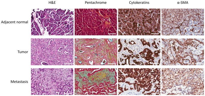
Experimental design: We sought to assess desmoplasia in metastatic lesions of PDAC and compare it with that of primary tumors. Fifty-three patients' primaries and 57 patients' metastases were stained using IHC staining techniques.
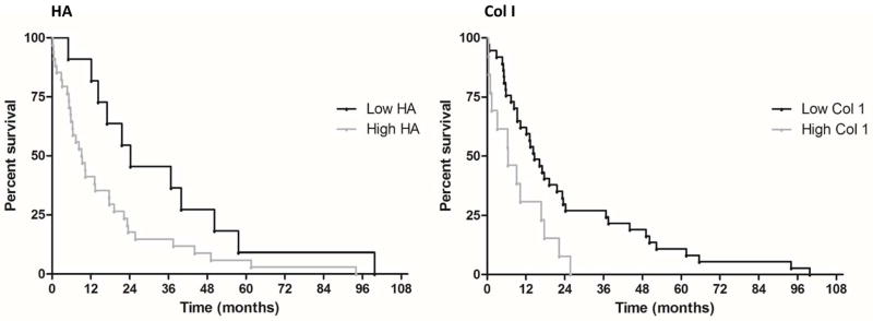
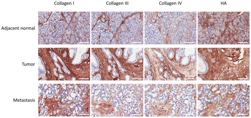
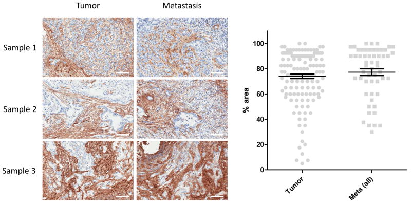
Results: We observed a significant negative correlation between patient survival and extracellular matrix deposition in primary tumors. Kaplan-Meier curves for collagen I showed median survival of 14.6 months in low collagen patients, and 6.4 months in high-level patients (log rank, P < 0.05). Low-level hyaluronan patients displayed median survival times of 24.3 months as compared with 9.3 months in high-level patients (log rank, P < 0.05). Our analysis also indicated that extracellular matrix components, such as collagen and hyaluronan, are found in high levels in both primary tumors and metastatic lesions. The difference in the level of desmoplasia between primary tumors and metastatic lesions was not statistically significant.
Conclusions: Our results suggest that both primary tumors and metastases of PDAC have highly fibrotic stroma. Thus, stromal targeting agents have the potential to benefit PDAC patients, even those with metastatic disease.
©2015 American Association for Cancer Research.
Conflict of interest statement
Potential conflict: PJ and HMS are employees of Halozyme Therapeutics. DDVH receives research support from Halozyme Therapeutics.
Figures
Comment in
-
Stroma, Stroma Everywhere (Far More Than You Think).Clin Cancer Res. 2015 Aug 1;21(15):3366-8. doi: 10.1158/1078-0432.CCR-15-0416. Epub 2015 May 15. Clin Cancer Res. 2015. PMID: 25979482 Free PMC article.
References
-
- Siegel R, Ma J, Zou Z, Jemal A. Cancer statistics. CA Cancer J Clin. 2014;64:9–29. - PubMed
-
- Mahadevan D, Von Hoff DD. Tumor-stroma interactions in pancreatic ductal adenocarcinoma. Mol Cancer Ther. 2007;6:1186–97. - PubMed
-
- Minchinton AI, Tannock IF. Drug penetration in solid tumours. Nat Rev Cancer. 2006;6:583–92. - PubMed
-
- Netti PA, Berk DA, Swartz MA, Grodzinsky AJ, Jain RK. Role of extracellular matrix assembly in interstitial transport in solid tumors. Cancer Res. 2000;60:2497–03. - PubMed
Publication types
MeSH terms
Substances
Grants and funding
LinkOut - more resources
Full Text Sources
Other Literature Sources
