. 2015 May;100(5):E710-9.
doi: 10.1210/jc.2014-4297. Epub 2015 Feb 19.

Pituitary adenoma with paraganglioma/pheochromocytoma (3PAs) and succinate dehydrogenase defects in humans and mice

Affiliations

Pituitary adenoma with paraganglioma/pheochromocytoma (3PAs) and succinate dehydrogenase defects in humans and mice

Paraskevi Xekouki et al. J Clin Endocrinol Metab. 2015 May.

Abstract

Context: Germline mutations in genes coding succinate dehydrogenase (SDH) subunits A, B, C, and D have been identified in familial paragangliomas (PGLs)/pheochromocytomas (PHEOs) and other tumors. We described a GH-secreting pituitary adenoma (PA) caused by SDHD mutation in a patient with familial PGLs. Additional patients with PAs and SDHx defects have since been reported.

Design: We studied 168 patients with unselected sporadic PA and with the association of PAs, PGLs, and/or pheochromocytomas, a condition we named the 3P association (3PAs) for SDHx germline mutations. We also studied the pituitary gland and hormonal profile of Sdhb(+/-) mice and their wild-type littermates at different ages.

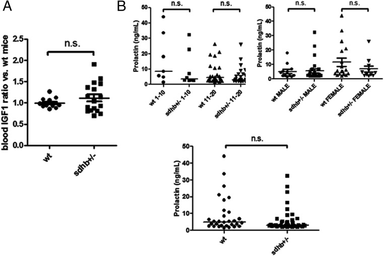
Results: No SDHx mutations were detected among sporadic PA, whereas three of four familial cases were positive for a mutation (75%). Most of the SDHx-deficient PAs were either prolactinomas or somatotropinomas. Pituitaries of Sdhb(+/-) mice older than 12 months had an increased number mainly of prolactin-secreting cells and several ultrastructural abnormalities such as intranuclear inclusions, altered chromatin nuclear pattern, and abnormal mitochondria. Igf-1 levels of mutant mice tended to be higher across age groups, whereas Prl and Gh levels varied according to age and sex.

Conclusion: The present study confirms the existence of a new association that we termed 3PAs. It is due mostly to germline SDHx defects, although sporadic cases of 3PAs without SDHx defects also exist. Using Sdhb(+/-) mice, we provide evidence that pituitary hyperplasia in SDHx-deficient cells may be the initial abnormality in the cascade of events leading to PA formation.


Figures

Figure 1.
Figure 1.
Hematoxylin and eosin-stained sections from pituitaries of 12-month-old WT (A and C) and Sdhb+/− (B and D) mice. Pituitaries of mutant mice appeared hyperplastic (D) compared with WT of the same age (C). Higher magnification revealed that pituitaries of mutant animals showed prominent dilated pools of extravasated blood cells (long arrows) (B) as well as intranuclear inclusions (E) (short arrows); the number of optic fields containing these inclusions was significantly higher in Sdhb+/− mice (n = 7 vs WT mice, n = 6) (P < .05, U test) (F).
Figure 2.
Figure 2.
Total number and proportion of Gh- and Prl-secreting cells pituitary cells per counting field in WT and Sdhb+/− mice. The total number of pituitary cells per optic field differed significantly between the two genotypes at 12 months old (P < .02) (A); an age-related decline was noted for both animal cohorts, which was nearly significant (P = .07, Student t test) for the WT mice (C). The number of Prl-positive cells was significantly higher in mutant mice at 12 months old (B); Gh-secreting cells tended to be higher at all ages in mutant mice (B). A decline in Gh- and Prl-secreting cells with age was also observed for both genotypes (C). Prl cells stained in red with Alexa Fluor 555 conjugated secondary antibody (D and E) and Gh-secreting cells green in immunofluorescent using Alexa Fluor 488-conjugated secondary antibody (F and G). Nuclei were stained with 4′,6′-diamino-2-phenylindole and in WT animals appeared oval shaped with central foci of condensed chromatin (H and J); in Sdhb+/− mice, loss of central foci of heterochromatin and accumulation adjacent to the nuclear membrane was noted (I and K).
Figure 3.
Figure 3.
Electron microscopic findings: adenohypophysis cells of the Sdhb+/− mice showed major morphological abnormalities of the mitochondria: many mitochondria were abnormally large in size (A) with marked swelling and abnormal and/or missing internal cristae (B and C) (arrows). These abnormalities were not seen in the WT animals (D). The intranuclear inclusions seen with the light microscopy appeared adjacent to the nucleus and presented like empty spaces (short arrows) (E and F). Staining for HIF-1α showed strong cytoplasmic HIF-1α expression (G) in pituitary of Sdhb+/− mouse compared with WT (H); a few cells demonstrated nuclear staining as an indication of hypoxia pathway activation (G, white arrows).
Figure 4.
Figure 4.
Igf-1 and Prl serum levels in WT and Sdhb+/− mice (median ± SE). A, Igf-1 levels in Sdhb+/− mice tended to be higher (1.3 times the levels of controls). Normalization is performed against the average hormone level measured in WT animals. B, Prl levels showed large variation, according to the age and sex of the animals. Upper left graph compares the Igf-1 levels between WT and Sdhb+/− mice grouped by age: 1–10 and 11–20 months old. Right panel compares the Igf-1 levels between WT and Sdhb+/− mice according to sex. Bottom panel compares WT vs Sdhb+/− mice with no distinction of age or sex. n.s., no significant difference by U tests (P > .05).

References

    1. Kantorovich V, King KS, Pacak K. SDH-related pheochromocytoma and paraganglioma. Best Pract Res Clin Endocrinol Metab. 2010;24:415–424. - PMC - PubMed
    1. Oyedotun KS, Lemire BD. The quaternary structure of the Saccharomyces cerevisiae succinate dehydrogenase. Homology modeling, cofactor docking, and molecular dynamics simulation studies. J Biol Chem. 2004;279:9424–9431. - PubMed
    1. Astuti D, Latif F, Dallol A, et al. Gene mutations in the succinate dehydrogenase subunit SDHB cause susceptibility to familial pheochromocytoma and to familial paraganglioma. Am J Hum Genet. 2001;69:49–54. - PMC - PubMed
    1. Baysal BE, Ferrell RE, Willett-Brozick JE, et al. Mutations in SDHD, a mitochondrial complex II gene, in hereditary paraganglioma. Science. 2000;287:848–851. - PubMed
    1. Burnichon N, Briere JJ, Libe R, et al. SDHA is a tumor suppressor gene causing paraganglioma. Hum Mol Genet. 2010;19:3011–3020. - PMC - PubMed

Publication types

Substances