Multicenter Study
. 2015 May 12;84(19):1948-55.
doi: 10.1212/WNL.0000000000001409. Epub 2015 Feb 18.

Prevalence and distribution of VZV in temporal arteries of patients with giant cell arteritis

Affiliations
Multicenter Study

Prevalence and distribution of VZV in temporal arteries of patients with giant cell arteritis

Don Gilden et al. Neurology. .

Abstract

Objective: Varicella-zoster virus (VZV) infection may trigger the inflammatory cascade that characterizes giant cell arteritis (GCA).

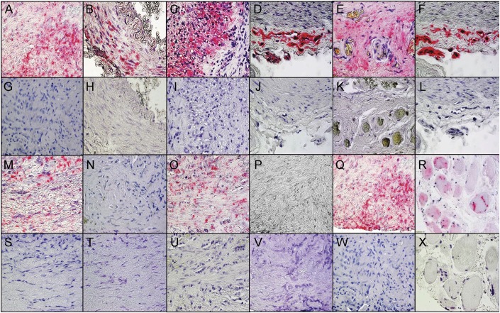
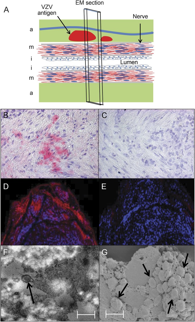
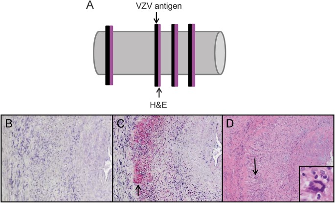
Methods: Formalin-fixed, paraffin-embedded GCA-positive temporal artery (TA) biopsies (50 sections/TA) including adjacent skeletal muscle and normal TAs obtained postmortem from subjects >50 years of age were examined by immunohistochemistry for presence and distribution of VZV antigen and by ultrastructural examination for virions. Adjacent regions were examined by hematoxylin & eosin staining. VZV antigen-positive slides were analyzed by PCR for VZV DNA.

Results: VZV antigen was found in 61/82 (74%) GCA-positive TAs compared with 1/13 (8%) normal TAs (p < 0.0001, relative risk 9.67, 95% confidence interval 1.46, 63.69). Most GCA-positive TAs contained viral antigen in skip areas. VZV antigen was present mostly in adventitia, followed by media and intima. VZV antigen was found in 12/32 (38%) skeletal muscles adjacent to VZV antigen-positive TAs. Despite formalin fixation, VZV DNA was detected in 18/45 (40%) GCA-positive VZV antigen-positive TAs, in 6/10 (60%) VZV antigen-positive skeletal muscles, and in one VZV antigen-positive normal TA. Varicella-zoster virions were found in a GCA-positive TA. In sections adjacent to those containing VZV, GCA pathology was seen in 89% of GCA-positive TAs but in none of 18 adjacent sections from normal TAs.

Conclusions: Most GCA-positive TAs contained VZV in skip areas that correlated with adjacent GCA pathology, supporting the hypothesis that VZV triggers GCA immunopathology. Antiviral treatment may confer additional benefit to patients with GCA treated with corticosteroids, although the optimal antiviral regimen remains to be determined.


Figures

Figure 1
Figure 1. VZV antigen in temporal arteries pathologically positive for GCA and in skeletal muscle adjacent to the infected temporal artery
Immunohistochemical analysis using mouse anti–varicella-zoster virus (VZV) glycoprotein E (gE) IgG1 antibody (see Methods) revealed VZV antigen in giant cell arteritis (GCA)-positive temporal arteries (TAs) in all arterial regions. VZV antigen (pink color) was seen in the adventitia and media (A), exclusively in the media (B), and in the media and intima (C) of 3 different GCA-positive TAs, as well as in the adventitia (D) of a positive control VZV-infected cadaveric cerebral artery 14 days after infection in vitro. The presence of VZV antigen was confirmed by immunostaining the same TA shown in panel A (E) and the same positive control VZV-infected cadaveric cerebral artery shown in panel D (F) with rabbit anti-VZV IE63 antibody. No staining was seen in sections adjacent to those containing VZV antigen when mouse isotype IgG1 antibody was substituted for mouse anti-VZV gE IgG1 antibody (G–J) or when normal rabbit serum was substituted for rabbit anti-VZV antibody (K, L). Discontinuous skip areas containing VZV antigen in a TA that was pathologically positive for GCA were shown by immunohistochemical staining with mouse anti-VZV gE IgG1 antibody as described in the Methods. VZV antigen was seen at positions 125–130 μm (M), 215–220 μm (O), and 255–260 μm (Q), but not at intervening regions at positions 131–214 μm (N) and 221–254 μm (P). Sections adjacent to those shown in panels M–Q were negative after immunostaining with mouse isotype IgG1 antibody (S–W). VZV antigen was often seen in skeletal muscle adjacent to a VZV-infected TA immunostained with mouse anti-VZV gE IgG1 antibody (R) that was not seen with mouse IgG1 antibody (X). 600× magnification.
Figure 2
Figure 2. Immunofluorescent staining and ultrastructural imaging of VZV-infected temporal artery
A giant cell arteritis–positive temporal artery (A) was examined for the presence of varicella-zoster virus (VZV) antigen in the adventitia (a), media (m), and intima (i). Immunohistochemical staining with rabbit anti-VZV IE63 antibody revealed VZV antigen in the media (B, pink color), but not after staining with rabbit anti–herpes simplex virus-1 antibody (C). Immunofluorescent staining with a different mouse anti-VZV IgG antibody than that used for immunohistochemistry in figure 1 revealed VZV antigen in the adventitia (D, red color), but not when primary antibody was omitted (E). Sections adjacent to those containing VZV antigen were prepared for transmission electron microscopy (TEM) and scanning electron microscopy (SEM) as described in the Methods. TEM revealed an enveloped virus particle (F, arrow), and SEM showed a cluster of virus particles in the adventitia egressing through an outer cell wall (G, arrows). Viral particles appear slightly larger than 200 nm because they were sputter coated with a gold alloy. In panels F and G, scale bars = 300 nm. EM = electron microscopy.
Figure 3
Figure 3. Pathologic analysis of sections adjacent to those containing VZV antigen from GCA-positive temporal arteries
Temporal arteries (TAs) in which varicella-zoster virus (VZV) antigen was detected immunohistochemically were further analyzed pathologically. Sections adjacent to those containing VZV antigen were stained by hematoxylin & eosin (H&E) (A). No staining was seen with mouse isotype IgG1 antibody (B). VZV antigen was found in nearly all giant cell arteritis (GCA)-positive TAs (C, pink color, arrow). H&E of the adjacent section showed classic GCA pathology (D), with inflammation, necrosis of the media, and giant cells (arrow, inset, 600× magnification) corresponding to VZV antigen (C, arrow). 100× magnification.

Comment in

References

    1. Lenton J, Donnelly R, Nash JR. Does temporal artery biopsy influence the management of temporal arteritis? QJM 2006;99:33–36. - PubMed
    1. Salazar R, Russman AN, Nagel MA, et al. Varicella zoster virus ischemic optic neuropathy and subclinical temporal artery involvement. Arch Neurol 2011;68:517–520. - PMC - PubMed
    1. Mathias M, Nagel MA, Khmeleva N, et al. VZV multifocal vasculopathy with ischemic optic neuropathy, acute retinal necrosis and temporal artery infection in the absence of zoster rash. J Neurol Sci 2013;325:180–182. - PMC - PubMed
    1. Nagel MA, Russman AN, Feit H, et al. VZV ischemic optic neuropathy and subclinical temporal artery infection without rash. Neurology 2013;80:220–222. - PMC - PubMed
    1. Nagel MA, Bennett JL, Khmeleva N, et al. Multifocal VZV vasculopathy with temporal artery infection mimics giant cell arteritis. Neurology 2013;80:2017–2021. - PMC - PubMed

Publication types