. 2015 Feb 18;7(275):275ra22.
doi: 10.1126/scitranslmed.aaa4963.

Rational development and characterization of humanized anti-EGFR variant III chimeric antigen receptor T cells for glioblastoma

Affiliations

Rational development and characterization of humanized anti-EGFR variant III chimeric antigen receptor T cells for glioblastoma

Laura A Johnson et al. Sci Transl Med. .

Abstract

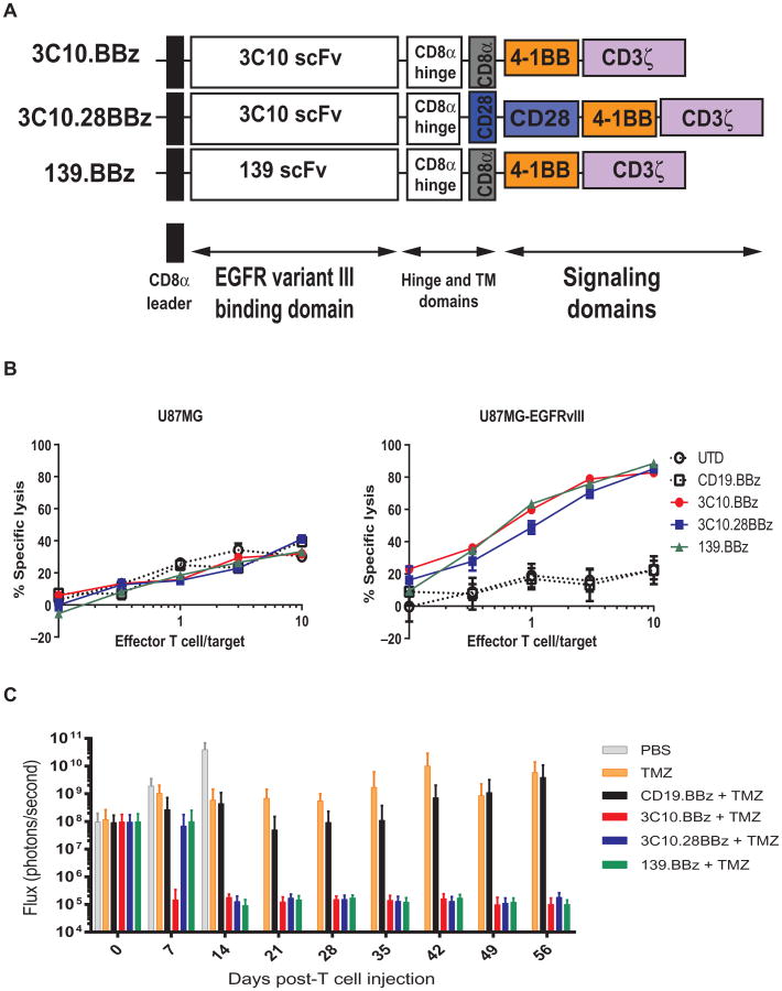
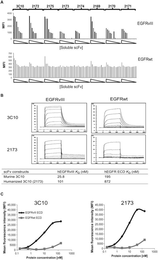
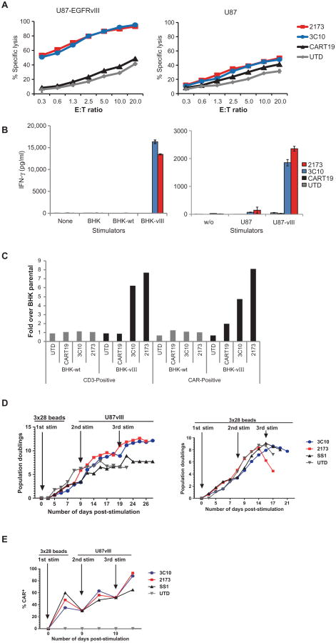
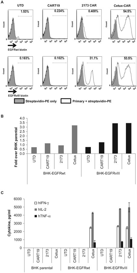
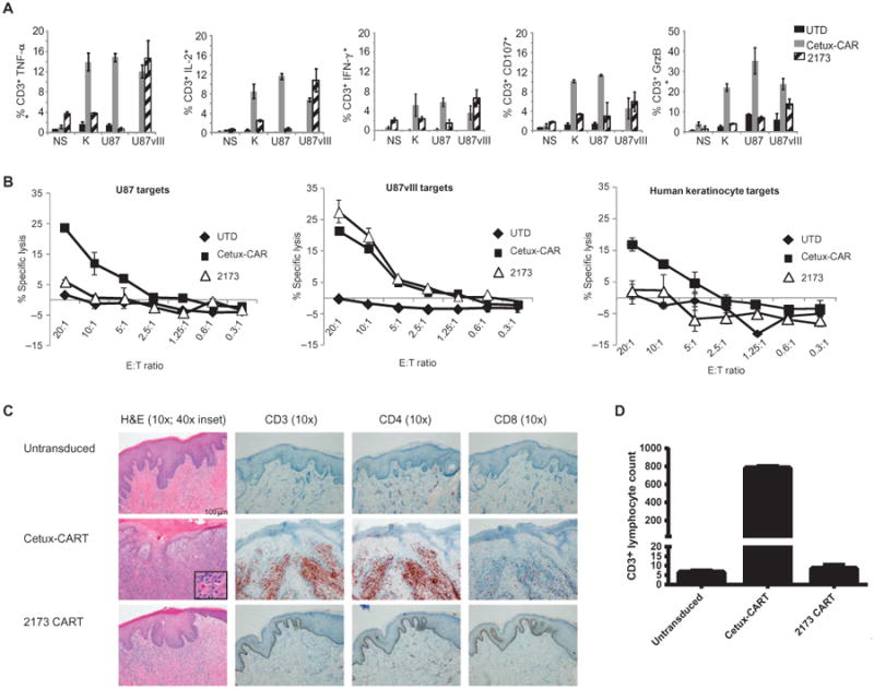
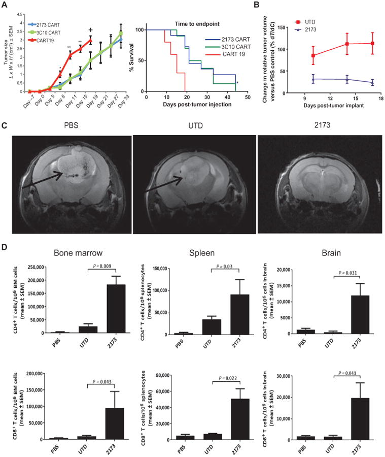
Chimeric antigen receptors (CARs) are synthetic molecules designed to redirect T cells to specific antigens. CAR-modified T cells can mediate long-term durable remissions in B cell malignancies, but expanding this platform to solid tumors requires the discovery of surface targets with limited expression in normal tissues. The variant III mutation of the epidermal growth factor receptor (EGFRvIII) results from an in-frame deletion of a portion of the extracellular domain, creating a neoepitope. We chose a vector backbone encoding a second-generation CAR based on efficacy of a murine scFv-based CAR in a xenograft model of glioblastoma. Next, we generated a panel of humanized scFvs and tested their specificity and function as soluble proteins and in the form of CAR-transduced T cells; a low-affinity scFv was selected on the basis of its specificity for EGFRvIII over wild-type EGFR. The lead candidate scFv was tested in vitro for its ability to direct CAR-transduced T cells to specifically lyse, proliferate, and secrete cytokines in response to antigen-bearing targets. We further evaluated the specificity of the lead CAR candidate in vitro against EGFR-expressing keratinocytes and in vivo in a model of mice grafted with normal human skin. EGFRvIII-directed CAR T cells were also able to control tumor growth in xenogeneic subcutaneous and orthotopic models of human EGFRvIII(+) glioblastoma. On the basis of these results, we have designed a phase 1 clinical study of CAR T cells transduced with humanized scFv directed to EGFRvIII in patients with either residual or recurrent glioblastoma (NCT02209376).


Figures

Fig. 1
Fig. 1. Selection of a 4-1BB-endodomain–based anti-EGFRvIII CAR construct
(A) Vector maps of tested anti-EGFRvIII CAR designs. (B) Flow-based cytotoxicity of CART-EGFRvIII cells against EGFRvIII+ glioma cell line. The glioblastoma cell line U87MG was stably transduced with EGFRvIII, fluorescently labeled with CFSE, and cocultured for 18 hours. Untransduced (UTD) or CD19.BBz CAR T cells were used as negative controls and compared to 3C10.BBz, 3C10.28BBz, or 139.BBz EGFRvIII CAR T cells. Lysis of parental cells (U87MG) and EGFRvIII-transduced U87 cells was analyzed at different effector/target (E:T) ratios by flow cytometry. One representative experiment is shown. Samples were performed in duplicate in two replicative experiments. (C) CART-EGFRvIII cells eradicate glioblastoma in an orthotopic xenogeneic mouse model. Seven days after 5 × 10⁴ U87-EGFRvIII cells were orthotopically implanted into mouse brains, mice were injected intravenously with either phosphate-buffered saline (PBS) alone, temozolomide (TMZ) alone, or temozolomide with 1 × 10⁶ transduced T cells expressing the indicated CAR constructs. Tumor burden was quantified as total flux in units of photons/second. Bars indicate means ± SD (n = 10 mice per group). Note that all mice in the PBS group died by day 15.
Fig. 2
Fig. 2. Selection of a humanized scFv with high specificity for EGFRvIII
(A) Binding of soluble scFvs to 293 cells transiently transfected with EGFRvIII or EGFRwt. Eight his-tagged solubilized humanized scFvs, at increasing concentrations from 0.002 to 200 nM, were incubated with either EGFRvIII-or EGFRwt-transfected 293 cells. After detection with phycoerythrin (PE)–conjugated anti-his secondary antibodies, the mean fluorescence intensity (MFI) was quantified. (B) Affinity measurements of soluble scFv binding to EGFRvIII or EGFRwt. Sensograms of soluble versions of the lead humanized 2173 scFv and the original murine 3C10 scFv flowed at various concentrations over a sensor chip binding the ECDs of EGFRvIII or EGFRwt. Association (after 240 s) or dissociation (after 360 s) rates were measured. Calculated KD values for these two scFvs for affinity of binding to either EGFRvIII or EGFRwt are shown in the table. (C) Membrane-bound CAR recognition of soluble EGFRvIII or EGFRwt recombinant proteins. MFI of murine 3C10– or humanized 2173 CAR–transduced T cells stained with varying concentrations of soluble and biotinylated forms of the ECDs of EGFRvIII or EGFRwt.
Fig. 3
Fig. 3. In vitro function of murine 3C10 and humanized 2173 CART-EGFRvIII cells
(A) Luciferase-based cytotoxicity assay of UTD, CART19, murine 3C10, or humanized 2173 CART-EGFRvIII cells cultured for 18 to 20 hours with U87 and U87-vIII target cells expressing luciferase. (B) Levels of interferon-γ (IFN-γ) as measured by enzyme-linked immunosorbent assay (ELISA) 24 hours after culturing UTD, CART19, murine 3C10, or humanized 2173 CART-EGFRvIII cells alone or with BHK, BHK-EGFRvIII (BHK-vIII), BHK-EGFRwt (BHK-wt), U87, or U87-EGFRvIII at a 1:1 E:T ratio. (C) Proliferation of UTD, CART19, 3C10, or 2173 CART-EGFRvIII cells after 6 days of coculture with either BHK-wt or BHK-vIII cells relative to proliferation over T cells cultured with parental BHK cells. (D) Long-term proliferation of T cells in response to repetitive stimulation with either antigen (U87-EGFRvIII cells) or anti-CD3/CD28 beads. T cells were either UTD or transduced to express either murine 3C10 or humanized EGFRvIII CAR, or an anti-mesothelin (SS1) CAR. All CARs are of the same vector design (4-1BB-ζ). (E) Enrichment of CAR-expressing T cells with repetitive antigen stimulation. CAR expression was quantified by flow cytometry just before each stimulation and normalized at each new stimulation as indicated by arrows.
Fig. 4
Fig. 4. In vitro comparison of humanized 2173 EGFRvIII-specific CAR to cetux-CAR
(A) Membrane-bound CAR recognition of soluble EGFRvIII or EGFR recombinant proteins. T cells that were either UTD or transduced with the indicated CARs were incubated with soluble biotinylated ECDs of EGFR or EGFRvIII. Histogram plots are shown with gates indicating the percentage of cells that stained with streptavidin-PE. (B) Proliferation of UTD or T cells transduced with the indicated CAR T cells after 4 days of coculture with BHK cells expressing EGFRwt or EGFRvIII. Y axis indicates fold proliferation over CAR T cells stimulated with parental BHK cells. (C) ELISA-based cytokine analysis on supernatants collected 24 hours after stimulation of T cells with BHK, BHK-EGFRwt, or BHK-EGFRvIII cells.
Fig. 5
Fig. 5. Evaluation of cross-reactivity of CAR T cells to human skin in vitro and in vivo
(A) Intracellular cytokine staining on untransduced (UTD), 2173 CAR, or cetux-CAR T cells after they were cocultured with no stimulation (NS), human keratinocytes (K), U87, or U87-EGFRvIII for 16 hours. Plots indicate percentage of T cells (±SD of two replicates) that stained positive for each cytokine as determined by flow cytometry. Data shown are representative of three independent experiments. GrzB, granzyme B. (B) Chromium release assays of cytotoxicity of UTD, 2173 CAR, or cetux-CAR T cells at ratios from 20:1 to 0.3:1 with either U87, U87-EGFRvIII, or human keratinocyte targets. Plots indicate means ± SEM of triplicate wells. (C) Hematoxylin and eosin (H&E)–stained human skin grafts excised from NSG mice 2 weeks after intravenous injection of either UTD, cetux-CAR–transduced, or humanized 2173 CAR–transduced T cells at ×10 magnification (degrees of magnification indicated at the top), along with an additional magnification inset in the cetux-CART–treated mice. On the right, immuno-histochemical stains for CD3, CD4, and CD8 of punch biopsy specimens obtained 3 days after T cell injection are shown. Scale bar, 100 mm (all 10× images). Results are representative of two experiments. (D) T cells infiltrating the epidermis were enumerated in a punch biopsy specimen. Bars show average and range of counts from two separate sections of adjacent cuts.
Fig. 6
Fig. 6. Antitumor activity of humanized 2173 CART-EGFRvIII cells in vivo
(A) NSG mice injected with U87-EGFRvIII tumors subcutaneously on day 0 and with T cells as indicated intravenously on day 7. Left plot indicates mean ± SEM of calculated tumor volume based on caliper measurements over time. Statistically significant differences between EGFRvIII CARs and CD19 CARs are marked by asterisks [*P = 0.01, **P = 0.003; n = 10 mice per group; one-way analysis of variance (ANOVA), Kruskal-Wallis test]. + indicates euthanasia. Survival based on time to endpoint was plotted using a Kaplan-Meier curve (Prism software); statistically significant differences between the experimental groups were determined using log-rank Mantel-Cox test (P = 0.0002). Endpoint was defined by tumor reaching 2 cm in any direction or a volume of 4 cm3, ulceration, more than 10% weight loss, or inability to ambulate. (B) Effect of 2173 CART-EGFRvIII cells and UTD T cells compared to PBS on intra cranial tumor volume over time. Mice were injected intracranially with 5 × 10⁴ U87-EGFRvIII cells on day 0; on day 7, mice were injected intravenously with 4 × 10⁶ CAR T cells or a matched number of total UTD T cells or PBS. Plots show means ± SEM of the calculated %TC for the days indicated. The study was terminated, and all mice were euthanized on day 18 due to the disease burden and condition of the mice in the PBS and UTD groups. n = 10 mice per group. (C) Magnetic resonance imaging (MRI) of intracranial U87-EGFRvIII tumors in mice 11 days after CAR T cell injection (day 18 of study). Arrows indicate tumor mass. n = 2 mice per group imaged with one representative image shown. (D) Numbers of CD4+ and CD8+ T cells in bone marrow (BM), spleen, and brain of intracranial tumor-bearing mice on day 18. Tissues were harvested, processed into a single-cell suspension, and stained with antibodies for enumeration of T cells by flow cytometry. Plots indicate means ± SEM number of T cells per million cells as measured by flow cytometry of each homogenized organ. Statistically significant differences were calculated by one-way ANOVA, using Dunnett's test (n = 8 mice per group; P values as indicated).

Comment in

Similar articles

Cited by

References

    1. Hodi FS, O'Day SJ, McDermott DF, Weber RW, Sosman JA, Haanen JB, Gonzalez R, Robert C, Schadendorf D, Hassel JC, Akerley W, van den Eertwegh AJ, Lutzky J, Lorigan P, Vaubel JM, Linette GP, Hogg D, Ottensmeier CH, Lebbe C, Peschel C, Quirt I, Clark JI, Wolchok JD, Weber JS, Tian J, Yellin MJ, Nichol GM, Hoos A, Urba WJ. Improved survival with ipilimumab in patients with metastatic melanoma. N Engl J Med. 2010;363:711–723. - PMC - PubMed
    1. Topalian SL, Hodi FS, Brahmer JR, Gettinger SN, Smith DC, McDermott DF, Powderly JD, Carvajal RD, Sosman JA, Atkins MB, Leming PD, Spigel DR, Antonia SJ, Horn L, Drake CG, Pardoll DM, Chen L, Sharfman WH, Anders RA, Taube JM, McMiller TL, Xu H, Korman AJ, Jure-Kunkel M, Agrawal S, McDonald D, Kollia GD, Gupta A, Wigginton JM, Sznol M. Safety, activity, and immune correlates of anti–PD-1 antibody in cancer. N Engl J Med. 2012;366:2443–2454. - PMC - PubMed
    1. Porter DL, Levine BL, Kalos M, Bagg A, June CH. Chimeric antigen receptor–modified T cells in chronic lymphoid leukemia. N Engl J Med. 2011;365:725–733. - PMC - PubMed
    1. Grupp SA, Kalos M, Barrett D, Aplenc R, Porter DL, Rheingold SR, Teachey DT, Chew A, Hauck B, Wright JF, Milone MC, Levine BL, June CH. Chimeric antigen receptor–modified T cells for acute lymphoid leukemia. N Engl J Med. 2013;368:1509–1518. - PMC - PubMed
    1. Maude SL, Frey N, Shaw PA, Aplenc R, Barrett DM, Bunin NJ, Chew A, Gonzalez VE, Zheng Z, Lacey SF, Mahnke YD, Melenhorst JJ, Rheingold SR, Shen A, Teachey DT, Levine BL, June CH, Porter DL, Grupp SA. Chimeric antigen receptor T cells for sustained remissions in leukemia. N Engl J Med. 2014;371:1507–1517. - PMC - PubMed

Publication types

Associated data