. 2015 May;21(5):452-65.
doi: 10.1093/molehr/gav007. Epub 2015 Feb 19.

Genome-wide DNA methylation identifies trophoblast invasion-related genes: Claudin-4 and Fucosyltransferase IV control mobility via altering matrix metalloproteinase activity

Affiliations

Genome-wide DNA methylation identifies trophoblast invasion-related genes: Claudin-4 and Fucosyltransferase IV control mobility via altering matrix metalloproteinase activity

Yuxiang Hu et al. Mol Hum Reprod. 2015 May.

Abstract

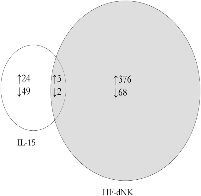
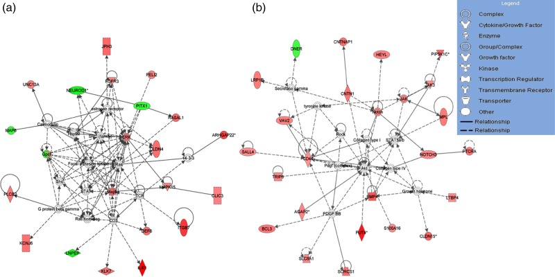
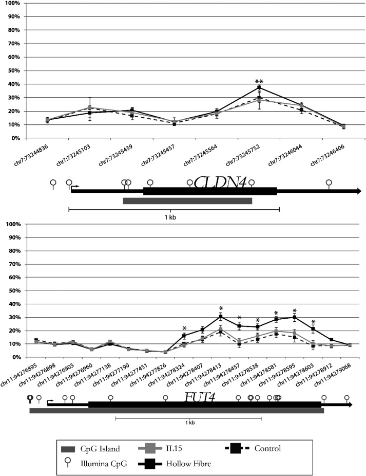
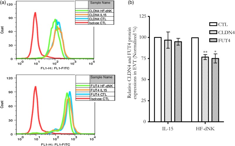
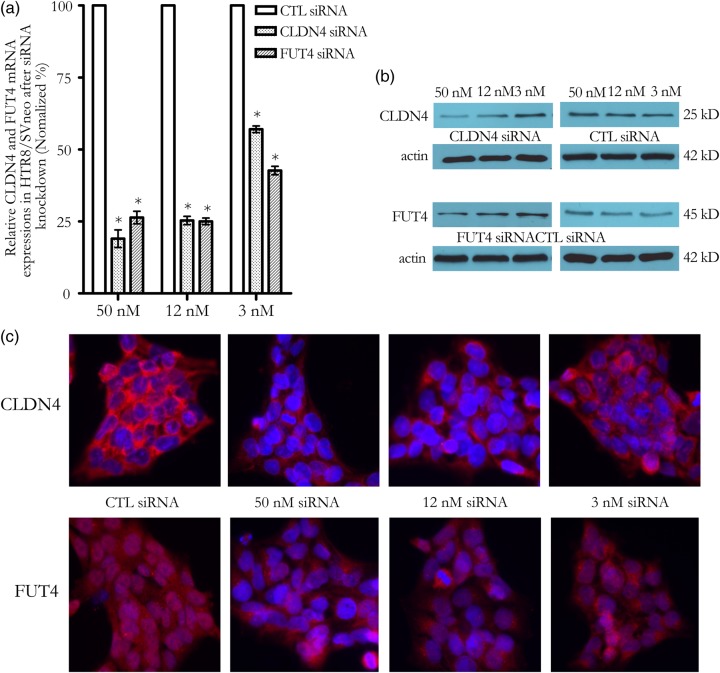
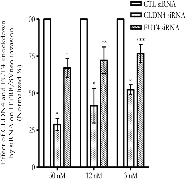
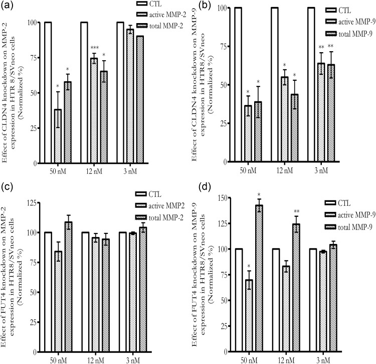
Previously we showed that extravillous cytotrophoblast (EVT) outgrowth and migration on a collagen gel explant model were affected by exposure to decidual natural killer cells (dNK). This study investigates the molecular causes behind this phenomenon. Genome wide DNA methylation of exposed and unexposed EVT was assessed using the Illumina Infinium HumanMethylation450 BeadChip array (450 K array). We identified 444 differentially methylated CpG loci in dNK-treated EVT compared with medium control (P < 0.05). The genes associated with these loci had critical biological roles in cellular development, cellular growth and proliferation, cell signaling, cellular assembly and organization by Ingenuity Pathway Analysis (IPA). Furthermore, 23 mobility-related genes were identified by IPA from dNK-treated EVT. Among these genes, CLDN4 (encoding claudin-4) and FUT4 (encoding fucosyltransferase IV) were chosen for follow-up studies because of their biological relevance from research on tumor cells. The results showed that the mRNA and protein expressions of both CLDN4 and FUT4 in dNK-treated EVT were significantly reduced compared with control (P < 0.01 for both CLDN4 and FUT4 mRNA expression; P < 0.001 for CLDN4 and P < 0.01 for FUT4 protein expression), and were inversely correlated with DNA methylation. Knocking down CLDN4 and FUT4 by small interfering RNA reduced trophoblast invasion, possibly through the altered matrix metalloproteinase (MMP)-2 and/or MMP-9 expression and activity. Taken together, dNK alter EVT mobility at least partially in association with an alteration of DNA methylation profile. Hypermethylation of CLDN4 and FUT4 reduces protein expression. CLDN4 and FUT4 are representative genes that participate in modulating trophoblast mobility.

Keywords: Claudin-4; DNA methylation; Fucosyltransferase IV; decidual natural killer cells; extravillous cytotrophoblast.


Figures

Figure 1
Figure 1
Venn diagram summary of differentially methylated loci from interleukin (IL)-15 and Hollow fiber-decidual natural killer cells (HF-dNK)-treated extravillous cytotrophoblast (EVT). HF-dNK treatment on EVT resulted in 444 candidate loci with methylation alteration compared with medium control, and among them, there were 5 loci in common with IL-15 treated EVT. Number of differentially methylated loci is indicated in the circles. ↑: hypermethylated; ↓: hypomethylated.
Figure 2
Figure 2
The two representative networks by ingenuity pathway analysis (IPA) for differentially methylated whole gene set derived from HF-dNK-treated EVT. (a) Extracellular signal-regulated protein kinase 1 and 2 (Erk1/2) was centered in one of the top network pathways, and Claudin-4 (CLDN4) was revolved this pathway. (b) Protein kinase B (AKT) was centered in another top network pathway, and fucosyltransferase IV (FUT4) was revolved this pathway. The analyses indicate that the differentially methylated genes from HF-dNK-treated EVT are located in pathways related to cellular development, cellular function and maintenance, and cell-to-cell signaling and interaction, revolving around key cellular proteins AKT and Erk1/2. The color intensity of node indicates the degree of hyper- or hypo-methylation. Genes encoding for molecules in red are hypermethylated while those in green are hypomethylated relative to control EVT. The shape of each node denotes the molecule class displayed beside each figure. The nodal relationships indicated in solid lines denote direct while those in dashed lines denote indirect interactions. AGAP2: ArfGAP with GTPase domain, ankyrin repeat and PH domain 2; ARC: Activity-regulated cytoskeleton-associated protein; BCL3: B cell lymphoma 3 protein; BMP4: bone morphogenetic protein 4; CD3: cluster of differentiation 3; CD44: cluster of differentiation 44; CLIC3: chloride intracellular channel 3; CNTN1: contactin 1; CNTNAP1: contactin associated protein 1; DEF6: differentially expressed in FDCP 6 homolog; DNER: delta and notch-like epidermal growth factor-related receptor; FGFR3: fibroblast growth factor receptor 3; GPC1: glypican 1; HEYL: hes-related family bHLH transcription factor with YRPW motif-like; Hsp90: 90 kDa heat shock protein; IL4I1: interleukin 4 induced 1; ITGB7: intergrin, beta 7; JAK: janus kinase; JAK3: janus kinase 3; KCNJ6: potassium inwardly-rectifying channel, subfamily J, member 6; KLK7: kallikrein-related peptidase 7; LNPEP: leucyl/cystinyl aminopeptidase; LRP1B: low density lipoprotein receptor-related protein 1B; MAP6: microtubule-associated protein 6; MAPK: mitogen-activated protein kinase; MPL: myeloproliferative leukemia virus oncogene; NEUROD1: neuronal differentiation 1; NOTCH3: Notch homolog 3; PDGF BB: platelet-derived growth factor beta polypeptide homodimer; PELI2: pellino E3 ubiquitin protein ligase family member 2; PIP5K1C: phosphatidylinositol-4-phosphate 5-kinase, type I, gamma; PITX1: paired-like homeodomain 1; PLCB2: phospholipase C, beta 2; PTCRA: pre T-cell antigen receptor alpha; RASAL1: RAS protein activator like 1; ROCK: rho-associated, coiled-coil-containing protein kinase; S100A16: S100 calcium binding protein A16; SALL4: spalt-like transcription factor 4; Shc: SHC (Src homology 2 domain containing) transforming protein; SLC8A1: solute carrier family 8 (sodium/calcium exchanger), member 1; SORCS1: sortilin-related VPS10 domain containing receptor 1; STAT: signal transducer and activator of transcription; STAT5a/b: signal transducer and activator of transcription 5a/b; TCR: T cell receptor; TRIP6: thyroid hormone receptor interactor 6; UNC13A: protein unc-13 homolog A; VAV2: vav 2 guanine nucleotide exchange factor.
Figure 3
Figure 3
CLDN4 and FUT4 methylation probe allocation in Illumina HumanMethylation450 array (450 array). CLDN4 and FUT4 DNA methylation probes in 450 array were targeting multiple CpG sites across whole gene regions. The CpG sites with significantly increased CpG methylation were located in the enhancer region (or first Exon). CLDN4 (chr7: 73245752); FUT4 (chr11: 94278324-chr11: 94278603) *P < 0.001; **P < 0.01.
Figure 4
Figure 4
CLDN4 and FUT4 protein expressions in EVT from explant culture. (a) CLDN4 and FUT4 protein expressions measured by flow cytometry; (b) mean density of flow cytometry for CLDN4 and FUT4. *P < 0.001; **P < 0.01. CLDN4 and FUT4 protein expressions were reduced by exposure to dNK-HF but not IL-15. CTL: control.
Figure 5
Figure 5
CLDN4 and FUT4 mRNA and protein expressions in HTR8/SVneo cells. (a) CLDN4 and FUT4 mRNA expressions were reduced by small interfering RNA (siRNA) transfection as measured by qPCR. *P < 0.001; (b and c) CLDN4 and FUT4 protein expressions were reduced by siRNA transfection as measured by western blot (b) and immunocytochemistry staining (c). CLDN4 displayed a primary membrane staining, and FUT4 was a primary cytoplasmic staining. Blue color showed the 4′,6-Diamidino-2-phenylindole (DAPI) nuclear staining; Red color was CLDN4 or FUT4 protein staining. Magnification of images: ×400.
Figure 6
Figure 6
Knockdown of CLDN4 and FUT4 expressions by siRNA in HTR8/SVneo reduces cell invasion. X-axis refers to the concentration of siRNA. *P < 0.001; **P < 0.01; ***P < 0.05.
Figure 7
Figure 7
Knockdown of CLDN4 and FUT4 expressions by siRNA in HTR8/SVneo alters matrix metalloproteinase (MMP)-2 and/or MMP-9 activities. (a) Knockdown CLDN4 decreased active MMP-2 and total MMP-2 activities; (b) Knockdown CLDN4 decreased active and total MMP-9 activities; (c) Knockdown FUT4 did not significantly alter active MMP-2 and total MMP-2 activities; (d) Knockdown FUT4 decreased active MMP-9 expression while increased total MMP-9 activity. X-axis refers to the concentrations of siRNA. *P < 0.001; **P < 0.01; ***P < 0.05.

Similar articles

Cited by

References

    1. Agarwal R, D'Souza T, Morin PJ. Claudin-3 and claudin-4 expression in ovarian epithelial cells enhances invasion and is associated with increased matrix metalloproteinase-2 activity. Cancer Res 2005;65:7378–7385. - PubMed
    1. Bertagnolo V, Benedusi M, Brugnoli F, Lanuti P, Marchisio M, Querzoli P, Capitani S. Phospholipase C-beta 2 promotes mitosis and migration of human breast cancer-derived cells. Carcinogenesis 2007;28:1638–1645. - PubMed
    1. Blair JD, Yuen RK, Lim BK, McFadden DE, von Dadelszen P, Robinson WP. Widespread DNA hypomethylation at gene enhancer regions in placentas associated with early-onset pre-eclampsia. Mol Hum Reprod 2013;19:697–708. - PMC - PubMed
    1. Boireau S, Buchert M, Samuel MS, Pannequin J, Ryan JL, Choquet A, Chapuis H, Rebillard X, Avances C, Ernst M, et al. DNA-methylation-dependent alterations of claudin-4 expression in human bladder carcinoma. Carcinogenesis 2007;28:246–258. - PubMed
    1. Braconi C, Huang N, Patel T. MicroRNA-dependent regulation of DNA methyltransferase-1 and tumor suppressor gene expression by interleukin-6 in human malignant cholangiocytes. Hepatology 2010;51:881–890. - PMC - PubMed

Publication types

MeSH terms