

Aggressive malignant phyllodes tumor
- PMID: 25697402
- PMCID: PMC4353966
- DOI: 10.1016/j.ijscr.2014.12.041
Aggressive malignant phyllodes tumor
Abstract
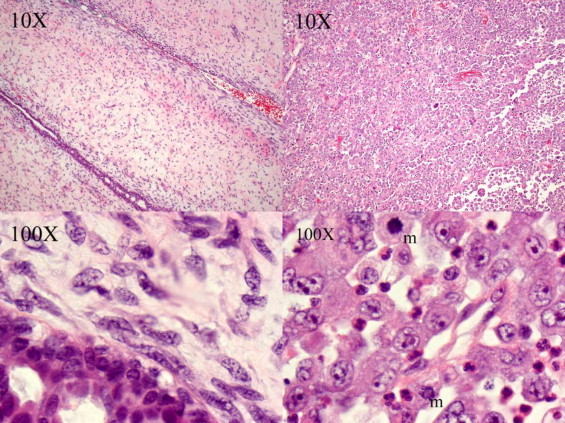
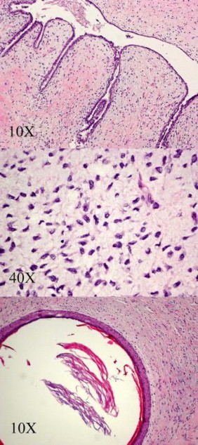
Introduction: Originally described in 1838 by Muller, phyllodes tumor is a rare fibroepithelial neoplasm which represents roughly 0.3-0.9% of all breast cancers. Phyllodes tumor are divided into benign, borderline and malignant histologic categories. Malignant phyllodes tumor represent anywhere from 10-30% of all phyllodes tumors. This group has both the potential to recur locally and metastasize, however not all malignant phyllodes behave this way. The challenge lays in predicting which tumor will recur locally or metastasize. Distinguishing this subset of malignant phyllodes tumor is paramount.
Presentation of case: We present a case of malignant phyllodes which presented with metastatic disease. What is fascinating about this case is not only the initial presentation but also the aggressiveness of this variation of phyllodes tumor. The patient initially presented with a large mass which encompassed her whole right breast. On surgical pathology the mass measured roughly 31cm in diameter and weighed over 10kg. Within 5 weeks from surgery the patient had suffered brain metastases and also 6 local recurrent tumors. The patient passed roughly 11 weeks after her first visit to our office.
Conclusion: Despite biopsy proven malignant phyllodes tumor, it was near impossible to predict such a rapid course of disease progression in our patient. Our case illustrates the unpredictable nature of this disease in general and it possibly sheds light on a variant of the disease which had undergone an aggressive transformation.
Keywords: Breast; Malignant; Phyllodes tumor.
Copyright © 2015 The Authors. Published by Elsevier Ltd.. All rights reserved.
Figures

References
-
- J. Muller, Uber den feineren Ban und Die Formen der Krankaften Geschwulste, about the ban and the finer forms of tumors krankaften Berlin, Germany: G Reimer (1838).
-
- Bernstein L., Deapen D. The descriptive epidemiology of malignant cystosarcoma phyllodes tumors of the breast. Cancer. 1993;71:3020–3024. - PubMed
-
- Azzopardi . Sarcoma in the breast. In: Benningron J., editor. Problems in Pathology. WB Saunders Co.; Philadelphia: 1979. pp. 355–359.
-
- Pietruszka B. Cystosarcoma phyllodes. a clinicopathological analysis of 42 cases. Cancer. 1978;41:1974–1983. - PubMed
LinkOut - more resources
Full Text Sources
Other Literature Sources
Research Materials
