. 2015 May 6:594:163-8.
doi: 10.1016/j.neulet.2015.01.084. Epub 2015 Feb 16.

The Fabry disease-associated lipid Lyso-Gb3 enhances voltage-gated calcium currents in sensory neurons and causes pain

Affiliations

The Fabry disease-associated lipid Lyso-Gb3 enhances voltage-gated calcium currents in sensory neurons and causes pain

L Choi et al. Neurosci Lett. .

Abstract

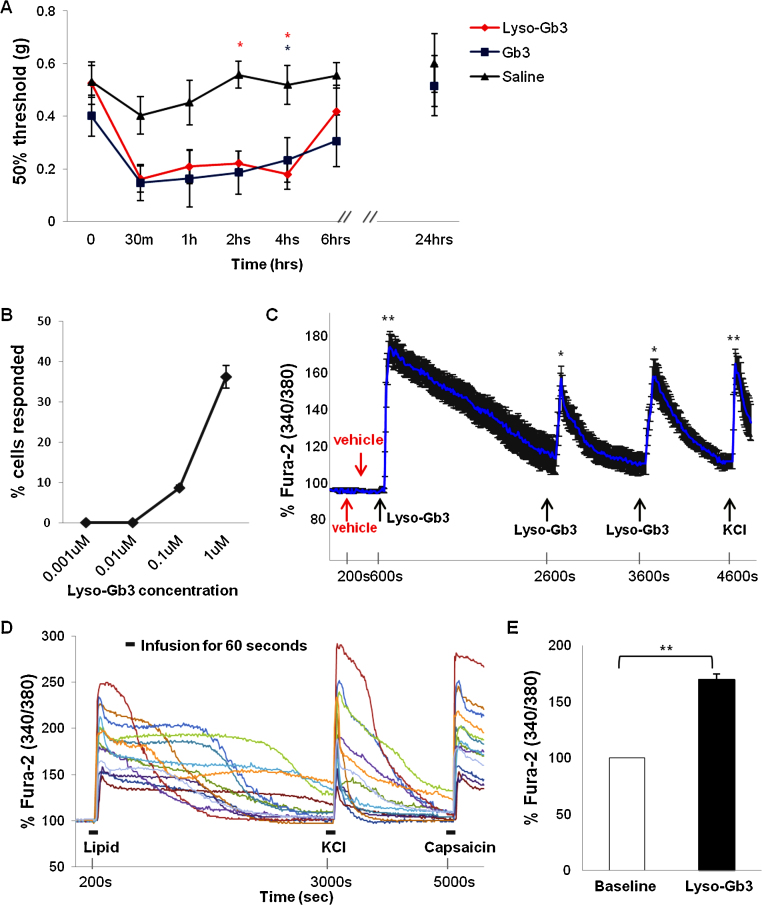
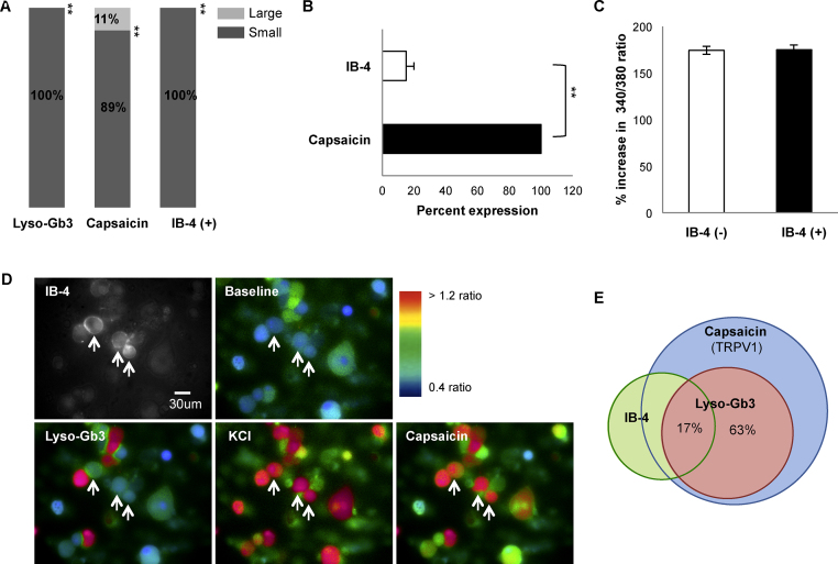
Fabry disease is an X-linked lysosomal storage disorder characterised by accumulation of glycosphingolipids, and accompanied by clinical manifestations, such as cardiac disorders, renal failure, pain and peripheral neuropathy. Globotriaosylsphingosine (lyso-Gb3), a deacylated form of globotriaosylceramide (Gb3), has emerged as a marker of Fabry disease. We investigated the link between Gb3, lyso-Gb3 and pain. Plantar administration of lyso-Gb3 or Gb3 caused mechanical allodynia in healthy mice. In vitro application of 100nM lyso-Gb3 caused uptake of extracellular calcium in 10% of sensory neurons expressing nociceptor markers, rising to 40% of neurons at 1μM, a concentration that may occur in Fabry disease patients. Peak current densities of voltage-dependent Ca(2+) channels were substantially enhanced by application of 1μM lyso-Gb3. These studies suggest a direct role for lyso-Gb3 in the sensitisation of peripheral nociceptive neurons that may provide an opportunity for therapeutic intervention in the treatment of Fabry disease-associated pain.

Keywords: Calcium imaging; Dorsal root ganglia; Fabry disease; Pain; Voltage-dependent Ca(2+) channels.


Figures

Fig. 1
Fig. 1
Effects of lyso-Gb3 on pain sensitivity and intracellular Ca2+ levels in nociceptive DRG neurons. (A) Either Gb3 or lyso-Gb3 injected into mice hind paws significantly enhanced pain sensitivity to mechanical stimuli over several hours post injection (n = 6–9 per group). Sensitivity resolved to a normal level within 24 h. Repeated measures ANOVA revealed effects of time: F (6,126) = 7.551, P < 0.0001, and of Fabry lipids (F (21,126) = 3.089, P < 0.001) but no difference between the two lipids (effect of lipid type: F (2,126) = 6.179, < 0.05). (B) Lyso-Gb3 evoked an increase in cytoplasmic Ca2+ levels in DRG neurons in a dose-dependent manner. The concentration of lyso-Gb3 producing changes in Ca2+ levels is plotted against the percentage of responsive DRG neurons. (C) Repetitive applications of lyso-Gb3 (1 μM), but not vehicle evoked transient increases in Ca2+ levels in DRG neurons. (D) Representative real time recordings of the changes in Fura-2 ratio acquired every 10 s in individual DRG neurons during bath applications of 1 μM Lyso-Gb3, 50 mM KCl and 1 μM capsaicin. (E) Statistical summary of the changes in Fura-2 ratio before and after application of 1 μM of lyso-Gb3 (n = 74). *P < 0.05 and **P < 0.0001.
Fig. 2
Fig. 2
Identification of the DRG neuron population responsive to lyso-Gb3. (A) Distribution of cells according to diameter; lyso-Gb3 responders were all small-diameter cells, as were 89 % of capsaicin-responsive cells. The third bar of the histogram signifies that IB-4 positive cells were all small-diameter neurons. (B) Lyso-Gb3 responsive cells were all stimulated by 1 μM capsaicin, but only 17 % of these bound IB-4.​ (B) Other lyso-Gb3 responsive capsaicin-sensitive cells were IB-4 negative. (C) Statistical summary of the lyso-Gb3-induced changes in Fura-2 ratio in neuronal subgroups shows no significant difference between IB4-positive and IB4-negative DRG neurons. (D) Representative images showing the changes in Fura-2 fluorescence during application of lyso-Gb3, KCl or capsaicin in IB4-positive DRG neurons. Arrows point to the IB4-positive neurons. (E) Subpopulation of lyso-Gb3-responding neurons represents approximately 63% of capsaicin-sensitive DRG neurons, only 17% of those were positive for IB-4 binding. **P < 0.0001.
Fig. 3
Fig. 3
Requirement for extracellular calcium in the Fura-2 response to lyso-GB3. (A) Representative real time traces showing intracellular Ca2+ levels following superfusion with lyso-Gb3 in either Ca2+ free or standard recording buffer containing 2 mM Ca2+. (B) Peak Fura-2AM 340/380 ratio between baseline and lyso-Gb3. Cells appear red in accordance with Ca2+ levels. (C) Representative fluorescent images showing changes in Ca2+ levels. Note: Data sampled every 10 s during calcium imaging. **P < 0.0001.
Fig. 4
Fig. 4
Effects of lyso-Gb3 on Ca2+ influx and current density of voltage-activated Ca2+ channels in DRG neurons. (A) Representative traces of Ca2+ currents evoked in the same small-diameter DRG neuron by voltage steps from holding potential (Vh) –70 mV to test potential (Vt) from –60 mV to +50 mV in 5 mV increments before (top) or during (medium) application of 1 μM lyso-Gb3 via a perfusion system. Ca2+ currents (bottom) in the presence of lyso-Gb3 were completely inhibited by 50 μM CdCl2. Inset illustrates the voltage-clamp protocol used to elicit currents. (B) Representative Ca2+ currents recorded at Vt 0 mV before (control) and after lyso-Gb3 application. (C) Average current–voltage relationships of Ca2+ currents before and during an application of 1 μM lyso-Gb3. The peak current amplitudes at each Vt were normalized to cell capacitance and expressed as the current density. (D) A statistical summary of the current densities at Vt 0 mV before and during lyso-Gb3 application. Changes in current density were normalized to control.. *P < 0.05 (paired t-test).

Comment in

References

    1. Fabry H. Angiokeratoma corporis diffusum–Fabry disease: historical review from the original description to the introduction of enzyme replacement therapy. Acta Paediatr. 2002;91:3–5. - PubMed
    1. Kaye E.M., Kolodny E.H., Logigian E.L., Ullman M.D. Nervous system involvement in Fabry’s disease: clinicopathological and biochemical correlation. Ann. Neurol. 1988;23:505–509. - PubMed
    1. Møller A.T., Jensen T.S. Neurological manifestations in Fabry’s disease. Nat. Clin. Pract. Neurol. 2007;3:95–106. - PubMed
    1. Dütsch M., Marthol H., Stemper B., Brys M., Haendl T., Hilz M.J. Small fiber dysfunction predominates in Fabry neuropathy. J. Clin. Neurophysiol. 2002;19:575–586. - PubMed
    1. Mills K., Morris P., Lee P., Vellodi A., Waldek S., Young E., Winchester B. Measurement of urinary CDH and CTH by tandem mass spectrometry in patients hemizygous and heterozygous for Fabry disease. J. Inherit. Metab. Dis. 2005;28:35–48. - PubMed

Publication types

MeSH terms