. 2015 Mar;110(2):14.
doi: 10.1007/s00395-015-0468-7. Epub 2015 Feb 20.

Loss of dihydrolipoyl succinyltransferase (DLST) leads to reduced resting heart rate in the zebrafish

Affiliations

Loss of dihydrolipoyl succinyltransferase (DLST) leads to reduced resting heart rate in the zebrafish

Mirjam Keßler et al. Basic Res Cardiol. 2015 Mar.

Abstract

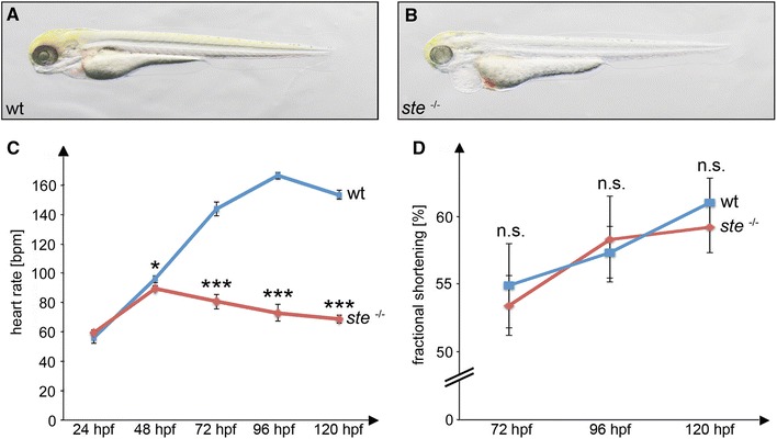
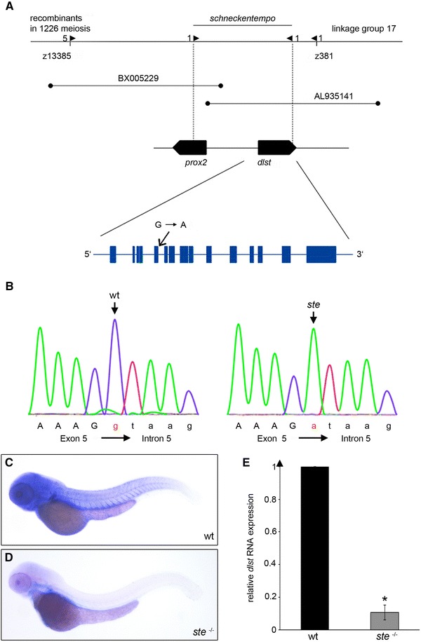
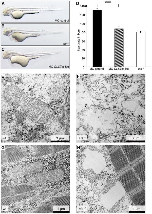
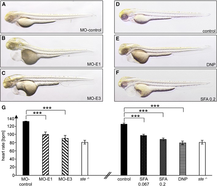
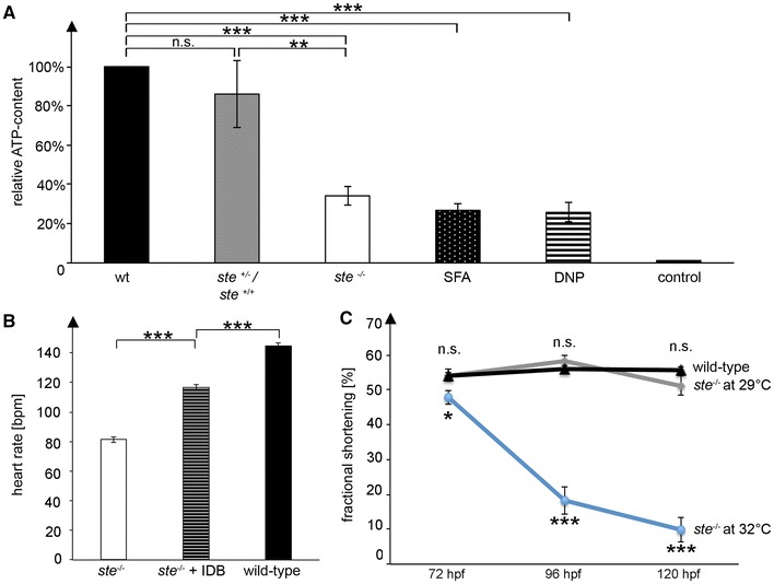
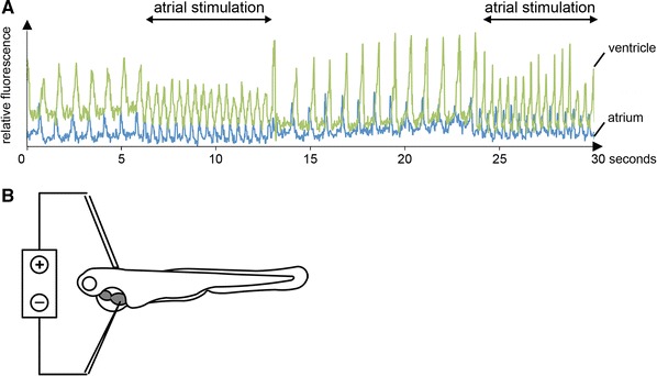
The genetic underpinnings of heart rate regulation are only poorly understood. In search for genetic regulators of cardiac pacemaker activity, we isolated in a large-scale mutagenesis screen the embryonic lethal, recessive zebrafish mutant schneckentempo (ste). Homozygous ste mutants exhibit a severely reduced resting heart rate with normal atrio-ventricular conduction and contractile function. External electrical pacing reveals that defective excitation generation in cardiac pacemaker cells underlies bradycardia in ste (-/-) mutants. By positional cloning and gene knock-down analysis we find that loss of dihydrolipoyl succinyltransferase (DLST) function causes the ste phenotype. The mitochondrial enzyme DLST is an essential player in the citric acid cycle that warrants proper adenosine-tri-phosphate (ATP) production. Accordingly, ATP levels are significantly diminished in ste (-/-) mutant embryos, suggesting that limited energy supply accounts for reduced cardiac pacemaker activity in ste (-/-) mutants. We demonstrate here for the first time that the mitochondrial enzyme DLST plays an essential role in the modulation of the vertebrate heart rate by controlling ATP production in the heart.


Figures

Fig. 1
Fig. 1
Effects of the ste mutation on cardiac function. a, b Lateral view of wild-type (wt) (a) and ste −/− mutant (b) embryos at 72 h post-fertilization (hpf). ste −/− mutants (b) develop blood congestion at the cardiac inflow tract and a pericardial edema. c Heart rate at different time points in ste −/− mutants (red) and wild-type littermates (blue). Bradycardia in ste −/− mutants starts at 48 hpf and becomes more severe during further embryonic development. d Ventricular fractional shortening is unaffected in ste −/− mutants compared to wild-type littermates at 72 hpf (53 ± 2.2 vs. 55 ± 3.1 %, n = 5, p = 0.7096), 96 hpf (58 ± 3.2 vs. 57 ± 1.9 %, n = 5, p = 0.7956) and 120 hpf (59 ± 1.9 vs. 61 ± 1.8 %, n = 5, p = 0.5586)
Fig. 2
Fig. 2
ste encodes for dihydrolipoyl succinyltransferase (dlst). a Integrated genetic and physical map of the ste locus. The ste mutation interval is flanked by the microsatellite markers z13385 and z381 and encodes two open reading frames, zebrafish dlst and prox2. b Within the coding sequence of zdlst a point mutation (G→A) was identified at the splice donor site of intron 5 leading to aberrant pre-mRNA splicing and the premature termination of DLST translation. An arrow marks the mutated base. c, d Spatial expression of dlst by whole-mount antisense RNA in situ hybridization of wt and ste −/− embryos at 72 hpf. c dlst is ubiquitously expressed with pronounced levels in the brain, skeletal muscle, fins and heart of wild-type embryos. d Strongly reduced dlst mRNA levels in ste −/− mutants compared to wild-type littermates. e Quantitative real-time PCR of ste −/− mutants and wild-type siblings. Nonsense mediated dlst-RNA decay in ste −/− mutants compared to wild-type siblings at 72 hpf
Fig. 3
Fig. 3
Targeted knockdown of zdlst phenocopies the ste −/− mutant phenotype. a, b Knockdown of zdlst by injection of Morpholino-modified antisense oligonucleotides (MO-DLSTsplice) (a) phenocopies the ste mutant phenotype (b), whereas injection of the same amount of standard control Morpholino (MO-control) (c) does not impact on heart rate. d Similar to ste −/− mutants, heart rate of DLSTsplice morphants is significantly reduced compared to MO-control-injected embryos at 72 hpf. e, f Transmission electron microscopy (TEM) of wild-type and ste −/− ventricular myocardium at 72 hpf. Scale bar represents 3 µM. e In wild-type myocardium, mitochondria display regular structure of inner and outer membrane and cristae. f In ste −/− myocardium, mitochondria display derangement of structure with severely reduced numbers of cristae. g, h TEM of wild-type and ste −/− skeletal muscle at 72 hpf. Scale bar represents 1 µM. g In wild-type skeletal muscle mitochondria exhibit regular morphology. h In ste −/− skeletal muscle mitochondria are characterized by large vacuoles and reduced numbers of cristae
Fig. 4
Fig. 4
Blockade of the citric acid cycle phenocopies the ste −/− mutant phenotype. ac, g Morpholino-modified antisense oligonucleotide mediated knockdown of OGDH subunit E1 (MO-E1) (a) or DLDH subunit E3 (MO-E3) (b) of the citric acid cycle phenocopies the ste −/− bradycardia (g), whereas injection of a control Morpholino has no effect (c, g). dg Incubation of wild-type embryos with the citric acid cycle blocker sodiumfluoroacetate (SFA) and the respiratory chain uncoupler 2,4-dinitrophenol (DNP) phenocopies the heart rate defect of ste −/− mutant embryos in a dose-dependent manner
Fig. 5
Fig. 5
Reduced ATP levels in ste −/− mutants lead to sinus bradycardia. a ATP content of ste −/− mutant embryos is significantly reduced compared to wild-type embryos (TE strain) and wild-type ste siblings (ste +/−;+/+ ), whereas ATP content between TE strain and wild-type ste siblings does not significantly differ. Dead zebrafish embryos serve as a control (72 hpf). The ATP content of embryos that were incubated with SFA and DNP is also significantly reduced and resembles the ATP content of ste −/− mutant embryos. b The coenzyme Q10 analogue Idebenone partially rescues heart rate in ste −/− mutant embryos that were incubated with this drug. c ste −/− mutants were incubated at increased temperature throughout embryonic development and ventricular fractional shortening was measured in wild-type and ste −/− mutant embryos at 72, 96 and 120 hpf. Fractional shortening of ste −/− mutant embryos under normal conditions (29 °C) does not significantly differ compared to wild-type embryos. Fractional shortening is reduced in ste −/− mutant embryos raised at 32 °C at (48 ± 2.1 vs. 54 ± 3.9 %, n = 10, p = 0.0358), 96 hpf (18 ± 4.0 vs. 56 ± 2.0 %, n = 10, p < 0.0001) and 120 hpf (10 ± 3.5 vs. 56 ± 2.1 %, n = 10, p < 0.0001)
Fig. 6
Fig. 6
a Selective atrial stimulation of ste −/− mutants at 72 hpf reconstitutes heart rate to normofrequency as depicted by simultaneous measurements of atrial (blue) and ventricular (green) cytoplasmic calcium transients. b Schematic description of the experimental setup for selective atrial stimulation using an electric stimulator

Similar articles

Cited by

References

    1. Abel ED, Doenst T. Mitochondrial adaptations to physiological vs. pathological cardiac hypertrophy. Cardiovasc Res. 2011;90:234–242. doi: 10.1093/cvr/cvr015. - DOI - PMC - PubMed
    1. Arrenberg AB, Stainier DY, Baier H, Huisken J. Optogenetic control of cardiac function. Science. 2010;330:971–974. doi: 10.1126/science.1195929. - DOI - PubMed
    1. Artuso L, Romano A, Verri T, Domenichini A, Argenton F, Santorelli FM, Petruzzella V. Mitochondrial DNA metabolism in early development of zebrafish (Danio rerio) Biochim Biophys Acta. 2012;1817:1002–1011. doi: 10.1016/j.bbabio.2012.03.019. - DOI - PubMed
    1. Benson DW, Wang DW, Dyment M, Knilans TK, Fish FA, Strieper MJ, Rhodes TH, George AL., Jr Congenital sick sinus syndrome caused by recessive mutations in the cardiac sodium channel gene (SCN5A) J Clin Invest. 2003;112:1019–1028. doi: 10.1172/JCI200318062. - DOI - PMC - PubMed
    1. Bonnefont JP, Chretien D, Rustin P, Robinson B, Vassault A, Aupetit J, Charpentier C, Rabier D, Saudubray JM, Munnich A. Alpha-ketoglutarate dehydrogenase deficiency presenting as congenital lactic acidosis. J Pediatr. 1992;121:255–258. doi: 10.1016/S0022-3476(05)81199-0. - DOI - PubMed

Publication types

Substances