. 2015 Jun:83:139-48.
doi: 10.1016/j.freeradbiomed.2015.01.035. Epub 2015 Feb 17.

MKK3 mediates inflammatory response through modulation of mitochondrial function

Affiliations

MKK3 mediates inflammatory response through modulation of mitochondrial function

Anup Srivastava et al. Free Radic Biol Med. 2015 Jun.

Abstract

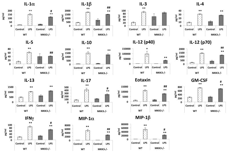
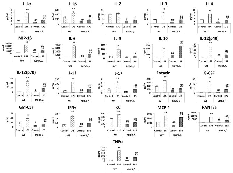
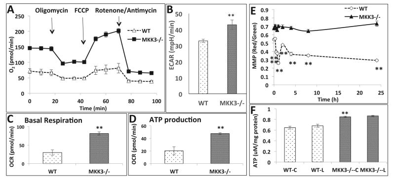
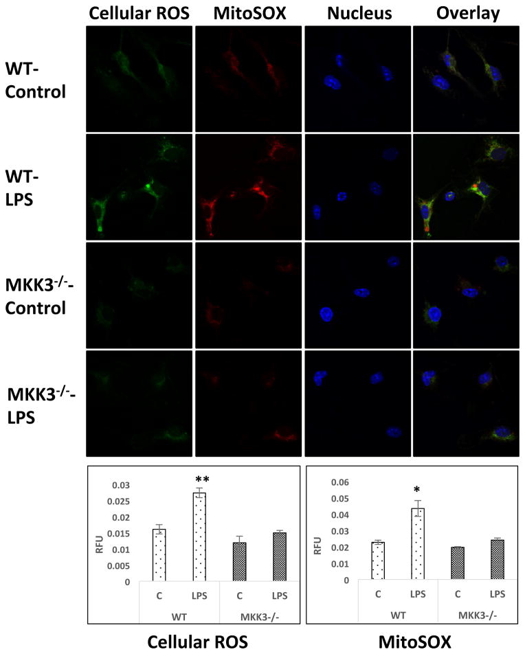
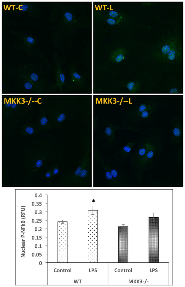
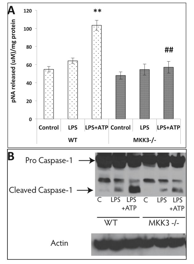
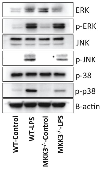
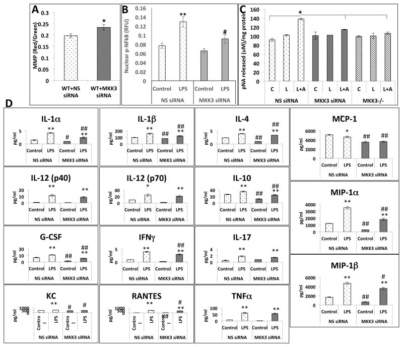
Mitochondria are increasingly recognized as drivers of inflammatory responses. MAP kinase kinase 3 (MKK3), a dual-specificity protein kinase, is activated in inflammation and in turn activates p38 MAP kinase signaling. Here we show that MKK3 influences mitochondrial function and acts as a critical mediator of inflammation. MKK3-deficient (MKK3(-/-)) mice and bone marrow-derived macrophages (BMDMs) secreted smaller amounts of cytokines than wild type (WT) after lipopolysaccharide (LPS) exposure. There was improved mitochondrial function, as measured by basal oxygen consumption rate, mitochondrial membrane potential, and ATP production, in MKK3(-/-) BMDMs. After LPS exposure, MKK3(-/-) BMDMs did not show a significant increase in cellular reactive oxygen species production or in mitochondrial superoxide compared to WT. Activation of two important inflammatory mediators, i.e., the nuclear translocation of NF-κB and caspase-1 activity (a key component of the inflammasome), was lower in MKK3(-/-) BMDMs. p38 and JNK activation was lower in MKK3(-/-) BMDMs compared to WT after exposure to LPS. Knockdown of MKK3 by siRNA in wild-type BMDMs improved mitochondrial membrane potential, reduced LPS-induced caspase-1 activation, and attenuated cytokine secretion. Our studies establish MKK3 as a regulator of mitochondrial function and inflammatory responses to LPS and suggest that MKK3 may be a therapeutic target in inflammatory disorders such as sepsis.

Keywords: Acute lung injury; Caspase 1; Free radicals; Inflammasome; Inflammation; Mitochondria; NF-κB; Oxidants; Sepsis.


Conflict of interest statement

Author Disclosure Statement

No conflicts of interest, financial or otherwise, are declared by the author(s).

Figures

Figure 1
Figure 1. Attenuated inflammatory responses in MKK3−/− mice against LPS induced injury
After 3h of LPS (40mg/kg) intra peritoneal injection in mice, serum was collected to analyze levels of cytokines. MKK3−/− mice showed attenuated inflammatory response compared to wild type mice after LPS injury. The baseline cytokine levels in MKK3−/− mice were not different than WT mice. * represents significance between control and LPS treated within same genotype and # represents significance between the genotypes with control and LPS treatment groups, * or # represents significance at p<0.05 and ** or ## represents significance at p<0.01. Measurements were done from serum of 3 mice per group.
Figure 2
Figure 2. Reduced inflammatory responses in MKK3−/− BMDMs against LPS stimulation
BMDM cell media was collected after 24h LPS (0.1μg/ml) treatment and cytokine levels were measured. MKK3−/− BMDMs showed lesser cytokine release after LPS treatment and also at baseline. * represents significance between control and LPS treated within same genotype and # represents significance between the genotypes with control and LPS treatment groups, * or # represents significance at p<0.05 and ** or ## represents significance at p<0.01. Data is representative of 3 independent experiments done in triplicates.
Figure 3
Figure 3. Improved mitochondrial bioenergetics in MKK3−/− BMDMs
Mitochondrial bioenergetic profile of BMDMs was studied using the “Mito stress kit”, Seahorse Biosciences. MKK3−/− BMDMs showed higher Oxygen consumption rate (OCR) compared to wild type (WT) (A). There was corresponding increase in extracellular acidification rate (ECAR) in MKK3−/− BMDMs (B). Basal respiration and ATP production was calculated based on the OCR profile and we found that they were higher in MKK3−/− BMDMs (C and D). The MMP and ATP were measured in MKK3−/− and WT BMDMs after LPS treatment. MMP as well as ATP content was higher in MKK3−/− BMDMs compared to WT at baseline and after LPS treatment MMP dropped in WT but not in MKK3−/− BMDMs (E and F). ** represents significance at p<0.01. Data is representative of 3 experiments done in at least 4 replicates.
Figure 4
Figure 4. Lower levels of reactive oxygen species (ROS) and mitochondrial superoxide in MKK3−/− BMDMs after LPS treatment
BMDMs were treated with LPS (0.1 μg/ml) for 60 min and stained with CM-H2DCFDA and MitoSOX. Cells were fixed and imaged with confocal microscopy and quantified using CellProfiler software. MKK3−/− BMDMs have lower levels of both cellular ROS as well as mitochondrial superoxide after LPS exposure. * and ** represents significance at p<0.05 and 0.01, respectively. Data is representative of atleast 20 images.
Figure 5
Figure 5. Lower NF-κB nuclear translocation in MKK3−/− BMDMs after LPS stimulation
BMDMs were treated with LPS (0.1 μg/ml) for 45 min and immuno-stained with p-NF-κB antibody. Slides where imaged with confocal microscopy and quantified using CellProfiler software. There was an increase in nuclear p-NF-κB in WT but to a lesser extent in MKK3−/− BMDMs compared to respective controls. * represents significance at p<0.05. Data is representative of 3 experiments done in at least duplicates.
Figure 6
Figure 6. Reduced caspase-1 activation in MKK3−/− BMDMs after LPS + ATP injury
BMDMs were treated with LPS and ATP to check the activation of caspase-1. Caspase-1 activity was significantly increased in WT after LPS (0.1 μg/ml) + ATP (5uM) treatment but MKK3−/− BMDMs did not show any significant change compared to control (A). The levels of cleaved caspase-1 were lower in MKK3−/− BMDMs compared to WT after LPS + ATP treatment (B). * represents significance between control and LPS treated within same genotype and # represents significance between the genotypes within LPS treatment groups, and ** or ## represents significance at p<0.01. Data is representative of 3 experiments done in at least triplicates.
Figure 7
Figure 7. MKK3−/− BMDMs show reduced activation of MAPK signaling after LPS stimulation
The MKK3−/− BMDMs after LPS (0.1 μg/ml) treatment showed lower levels of p-p38 and p-JNK compared to WT. There was no difference in p-ERK response after LPS treatment in both MKK3−/− and WT BMDMs. Data was normalized to b-actin content. Data is representative of 3 experiments done in at least triplicates with multiple western blotting runs.
Figure 8
Figure 8. MKK3 siRNA produces MKK3−/− phenotype in WT BMDMs
WT BMDMs transfected with MKK3 siRNA show higher MMP compared to control i.e. non-specific siRNA (NS siRNA) transfected (A). NF-κB nuclear translocation was lower in MKK3 siRNA transfected BMDMs compared to control after LPS treatment (0.1 μg/ml) (B). Caspase-1 activation was lower in MKK3 siRNA transfected BMDMs after LPS (0.1 μg/ml) + ATP (5 μM) injury compared to control (C). Cytokine release was lower in LPS (0.1 μg/ml) exposed MKK3 siRNA treated BMDMs compared to NS siRNA treamtment (D). For MMP graph * represents significance at p<0.05. For Caspase-1 activity and NF-κB graph * represents significance among treatments within same genotype at p<0.05. For cytokine graphs * represents significance between control and LPS treated within same siRNA treatment and # represents significance between different siRNA treatment with control and LPS treatment groups, * or # represents significance at p<0.05 and ** or ## represents significance at p<0.01. Data is representative of 3 experiments done in at least triplicates.

Similar articles

Cited by

References

    1. Lee I, Huttemann M. Energy crisis: The role of oxidative phosphorylation in acute inflammation and sepsis. Biochimica et biophysica acta. 2014;1842:1579–1586. - PMC - PubMed
    1. Huttemann M, Helling S, Sanderson TH, Sinkler C, Samavati L, Mahapatra G, Varughese A, Lu G, Liu J, Ramzan R, Vogt S, Grossman LI, Doan JW, Marcus K, Lee I. Regulation of mitochondrial respiration and apoptosis through cell signaling: cytochrome c oxidase and cytochrome c in ischemia/reperfusion injury and inflammation. Biochimica et biophysica acta. 2012;1817:598–609. - PMC - PubMed
    1. Debelec-Butuner B, Alapinar C, Varisli L, Erbaykent-Tepedelen B, Hamid SM, Gonen-Korkmaz C, Korkmaz KS. Inflammation-mediated abrogation of androgen signaling: an in vitro model of prostate cell inflammation. Molecular carcinogenesis. 2014;53:85–97. - PubMed
    1. Andrades ME, Ritter C, Dal-Pizzol F. The role of free radicals in sepsis development. Frontiers in bioscience. 2009;1:277–287. - PubMed
    1. Ryu MJ, Kim SJ, Kim YK, Choi MJ, Tadi S, Lee MH, Lee SE, Chung HK, Jung SB, Kim HJ, Jo YS, Kim KS, Lee SH, Kim JM, Kweon GR, Park KC, Lee JU, Kong YY, Lee CH, Chung J, Shong M. Crif1 deficiency reduces adipose OXPHOS capacity and triggers inflammation and insulin resistance in mice. PLoS genetics. 2013;9:e1003356. - PMC - PubMed

Publication types

MeSH terms

LinkOut - more resources