Review
. 2015 Apr 10;427(7):1589-608.
doi: 10.1016/j.jmb.2015.02.011. Epub 2015 Feb 16.

BiP and its nucleotide exchange factors Grp170 and Sil1: mechanisms of action and biological functions

Affiliations
Review

BiP and its nucleotide exchange factors Grp170 and Sil1: mechanisms of action and biological functions

Julia Behnke et al. J Mol Biol. .

Abstract

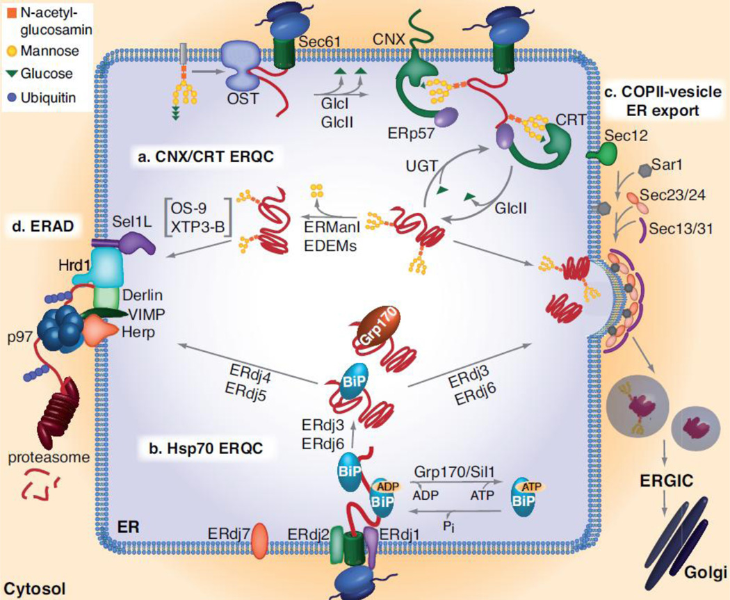
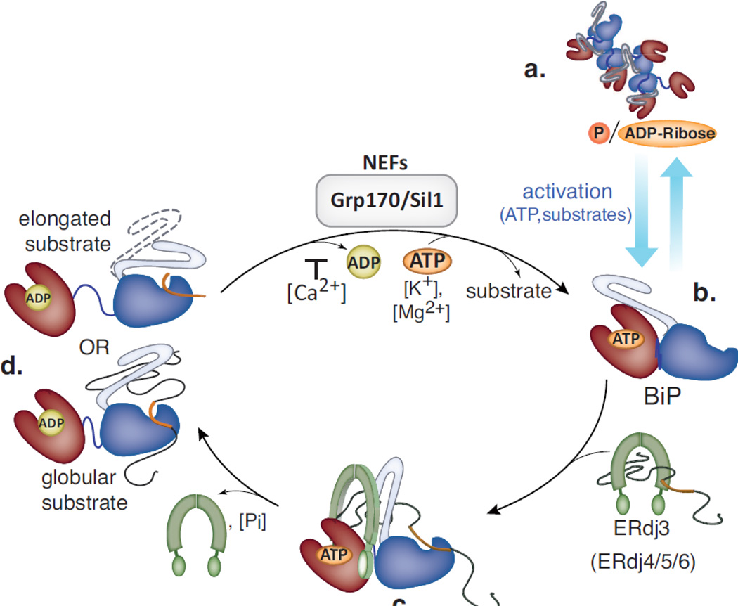
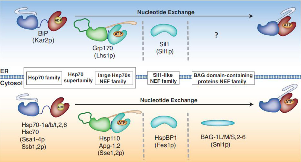
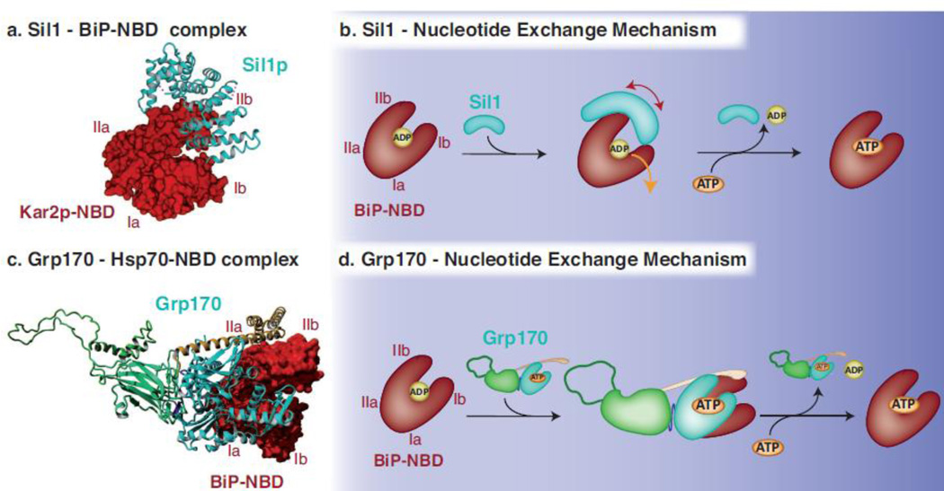
BiP (immunoglobulin heavy-chain binding protein) is the endoplasmic reticulum (ER) orthologue of the Hsp70 family of molecular chaperones and is intricately involved in most functions of this organelle through its interactions with a variety of substrates and regulatory proteins. Like all Hsp70 family members, the ability of BiP to bind and release unfolded proteins is tightly regulated by a cycle of ATP binding, hydrolysis, and nucleotide exchange. As a characteristic of the Hsp70 family, multiple DnaJ-like co-factors can target substrates to BiP and stimulate its ATPase activity to stabilize the binding of BiP to substrates. However, only in the past decade have nucleotide exchange factors for BiP been identified, which has shed light not only on the mechanism of BiP-assisted folding in the ER but also on Hsp70 family members that reside throughout the cell. We will review the current understanding of the ATPase cycle of BiP in the unique environment of the ER and how it is regulated by the nucleotide exchange factors, Grp170 (glucose-regulated protein of 170kDa) and Sil1, both of which perform unanticipated roles in various biological functions and disease states.

Keywords: BiP; Grp170; Sil1; nucleotide exchange factors; protein folding.


Conflict of interest statement

Conflict of interest statement

The authors declare no competing financial interests.

Figures

Fig. 1
Fig. 1. The ER Quality Control (ERQC) Machinery
Two main chaperone systems, the lectins CNX/CRT (a) and the Hsp70 chaperone BiP (b), aid the folding proteins for secretion (c) or if folding fails, target them for ERAD (d). (a) The oligosaccharyl transfer (OST) complex attaches a core oligosaccharide from a dolichol donor to the Asn of the Asn-X-Ser/Thr motif on nascent proteins during their translocation into the ER. GlcI and II remove the outer two glucose residues of the oligosaccharide, allowing the remaining glucose to be recognized by CNX/CRT. CNX/CRT assists protein folding in concert with further co-chaperones such as the protein-disulfide isomerase ERp57. Proteins exit the CNX/CRT cycle once the last glucose residue is removed by GlcII. If folded properly, the protein is released from the lectin chaperone cycle and is transported further along the secretory pathway. Incompletely folded intermediates can re-enter the CNX/CRT cycle if a single glucose is re-attached by the folding sensor UGT. If folding ultimately fails, proteins are further trimmed by ERManI and/or an EDEM resulting in removal of 4 mannose residues and recognition by OS-9 and XTP3-B, which then transfer the trimmed glycoprotein to the ERAD machinery for disposal. (b) The Hsp70 chaperone BiP binds hydrophobic patches exposed on nascent or incompletely folded proteins that are often non-glycosylated. BiP possesses low substrate binding affinity in the ATP-bound state and high affinity upon hydrolysis of ATP to ADP. Grp170 and Sil1 facilitate substrate release from BiP by stimulating the release of ADP and allowing ATP to rebind and open the lid on the substrate binding domain. Seven ERdj co-factors have been identified that interact with BiP via their J-domain and assist BiP in its functions during protein translocation (ERdj2), protein folding (ERdj3 and 6) and ERAD (ERdj4 and 5). The functions of ERdj1 and ERdj7 are not well understood, nor is the role of the large Hsp70, Grp170, that also binds to some incompletely folded BiP client proteins. (c) Once the threshold of folding set by the ERQC is met, proteins exit the ER in COPII-coated vesicles, a process that is initiated by Sec12 and driven by a GTPase, Sar1, and four major coat proteins, Sec23, Sec24, Sec13 and Sec31. (d) Once proteins that are clients of either chaperone system are delivered to the ERAD machinery, their retrotranslocation into the cytosol is facilitated by a complex of several transmembrane proteins including Sel1, Derlins, VIMP, Herp and Hrd1, which connect the machinery in the ER lumen to the protein ubiquitination machinery in the cytosol, allowing the ERAD client to be recognized by the p97 hexameric ATPase in the cytosol that provides the energy for extracting a protein from the ER for degradation by the 26S proteasome.
Fig. 2
Fig. 2. The ATPase cycle of BiP in the ER
(a) In the absence of high substrate loads, BiP exists in a multimeric form that is post-translationally modified (ADP-ribosylated and perhaps phosphorylated), which renders the protein inactive. When the demand for BiP increases, the modifications are removed allowing a readily accessable pool of BiP to be reactivated. (b). Once BiP binds potassium and ATP, its NBD (red) and its SBD (blue) come into close proximity to each other and the lid of the SBD (grey) opens, which in combination results in a form that binds substrates with low affinity. (c) Substrates can be introduced into the BiP cycle via their initial binding to DnaJ-like co-chaperones such as ERdj3 (green), which transfers substrate to BiP and increases BiP’s ATPase activity thereby locking the substrate onto BiP. Note that the binding sites within the substrate for ERdj3 and BiP (shown in orange) are probably not identical allowing a transient 3-way complex, which has been detected both in vivo and in vitro. (d) After the magnesium-dependent hydrolysis of ATP, BiP enters a state with low on and off rates for substrates. For elongated/peptide substrates the lid closes over the bound substrate, whereas for globular substrates direct interactions between the lid and the substrate exist but the lid may not close completely. The SBD and NBD become more distant upon substrate binding and ATP hydrolysis, which is less pronounced for globular substrates. To release the substrate and make BiP available for another round of client binding, ADP has to be exchanged against ATP. Calcium increases the affinity for ADP, whereas the NEFs Grp170 and Sil1 facilitate the nucleotide exchange reaction.
Fig. 3
Fig. 3. ER resident and cytosolic Hsp70 family members and their Nucleotide Exchange Factors
Several members of the Hsp70 protein family exist in the cytosol (bottom) of eukaryotic cells (yeast homologues are indicated in parentheses), whereas a single member, BiP resides in the ER (top). Three different classes of Hsp70 NEFs have been identified in the cytosol: large Hsp70 family members, Sil1-like and BAG-domain proteins that remove ADP from the NBD of Hsp70s, thus allowing ATP to bind and substrates to be released. A single member of the first and second class of NEFs, Grp170 and Sil1 respectively, have been found to act as nucleotide exchange factors for BiP in the ER. While Sil1-like and BAG-domain containing proteins are structurally unrelated to Hsp70 proteins, the large Hsp70 family of NEFs share many structural features with conventional Hsp70. The Hsp70 superfamily comprises conventional as well as large Hsp70s.
Fig. 4
Fig. 4. Mechanisms used by Sil1 and Grp170 to regulate nucleotide exchange for BiP
(a) The crystal structure obtained for yeast Sil1 bound to the NBD of BiP (PDB: 3QML) depicts how the single domain NEF Sil1, consisting of four Armadillo-like repeats (cyan, shown in ribbon), wraps around lobe IIb of BiP’s NBD (red, shown in surface representation) and displaces lobe Ib during the nucleotide exchange reaction. (b) Upon the binding of Sil1 to the ADP-bound form of BiP, the NBD cleft of BiP is opened by tilting lobe IIb and to a lesser extent lobe Ib outwards and thus destabilizing the domain and releasing ADP. ATP can subsequently bind to the NBD and BiP can re-enter its ATPase cycle. (c) The crystal structure of yeast Sse1p bound to the NBD of human Hsp70 (PDB: 3D2F) was used to model Grp170 (NBD in cyan, β-sheet and unstructured loop in green and α-helical domain in yellow, shown in ribbon) using Yasara Structure (www.yasara.org). Grp170 is shown bound to the human BiP NBD (3LDL) (red, shown in surface representation). Complex formation occurs via multiple contacts between the respective NBDs, and in addition, the C-terminal α-helical domain of Grp170 reaches out to embrace the Hsp70 NBD. (d) For the nucleotide exchange reaction to occur, Grp170 apparently binds to the ADP-bound form of BiP and destabilizes the structure of BiP´s NBD resulting in the release of ADP. Once ATP is bound to BiP, the Grp170-BiP complex would dissociate and BiP can re-enter its ATPase cycle, although key steps in the NEF activity for large Hsp70 proteins remain to be elucidated.

Similar articles

Cited by

References

    1. Hammond C, Braakman I, Helenius A. Role of N-linked oligosaccharide recognition, glucose trimming, and calnexin in glycoprotein folding and quality control. Proc Natl Acad Sci U S A. 1994;91:913–917. - PMC - PubMed
    1. Molinari M, Helenius A. Chaperone selection during glycoprotein translocation into the endoplasmic reticulum. Science. 2000;288:331–333. - PubMed
    1. Melnyk A, Rieger H, Zimmermann R. Co-chaperones of the Mammalian Endoplasmic Reticulum. Subcell Biochem. 2015;78:179–200. - PubMed
    1. Otero JH, Lizak B, Hendershot LM. Life and death of a BiP substrate. Semin Cell Dev Biol. 2010;21:472–478. - PMC - PubMed
    1. Chung KT, Shen Y, Hendershot LM. BAP, a mammalian BiP-associated protein, is a nucleotide exchange factor that regulates the ATPase activity of BiP. J Biol Chem. 2002;277:47557–47563. - PubMed

Publication types

MeSH terms