

Measuring the effects of aging and sex on regional brain stiffness with MR elastography in healthy older adults
- PMID: 25698157
- PMCID: PMC4387012
- DOI: 10.1016/j.neuroimage.2015.02.016
Measuring the effects of aging and sex on regional brain stiffness with MR elastography in healthy older adults
Abstract
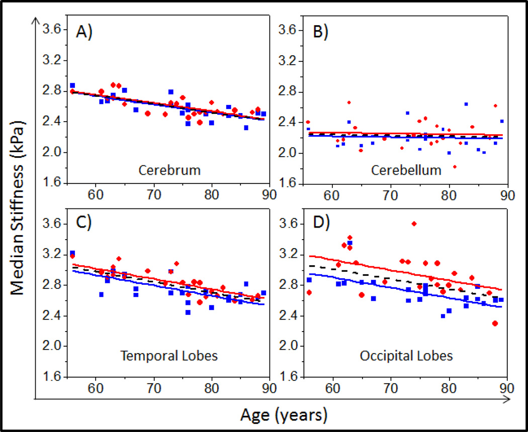
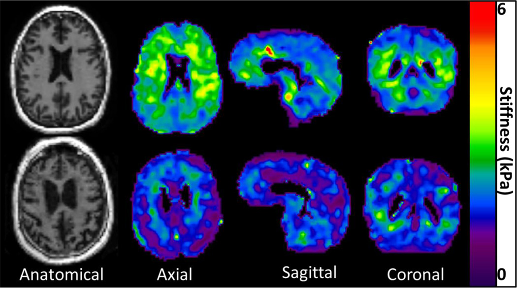
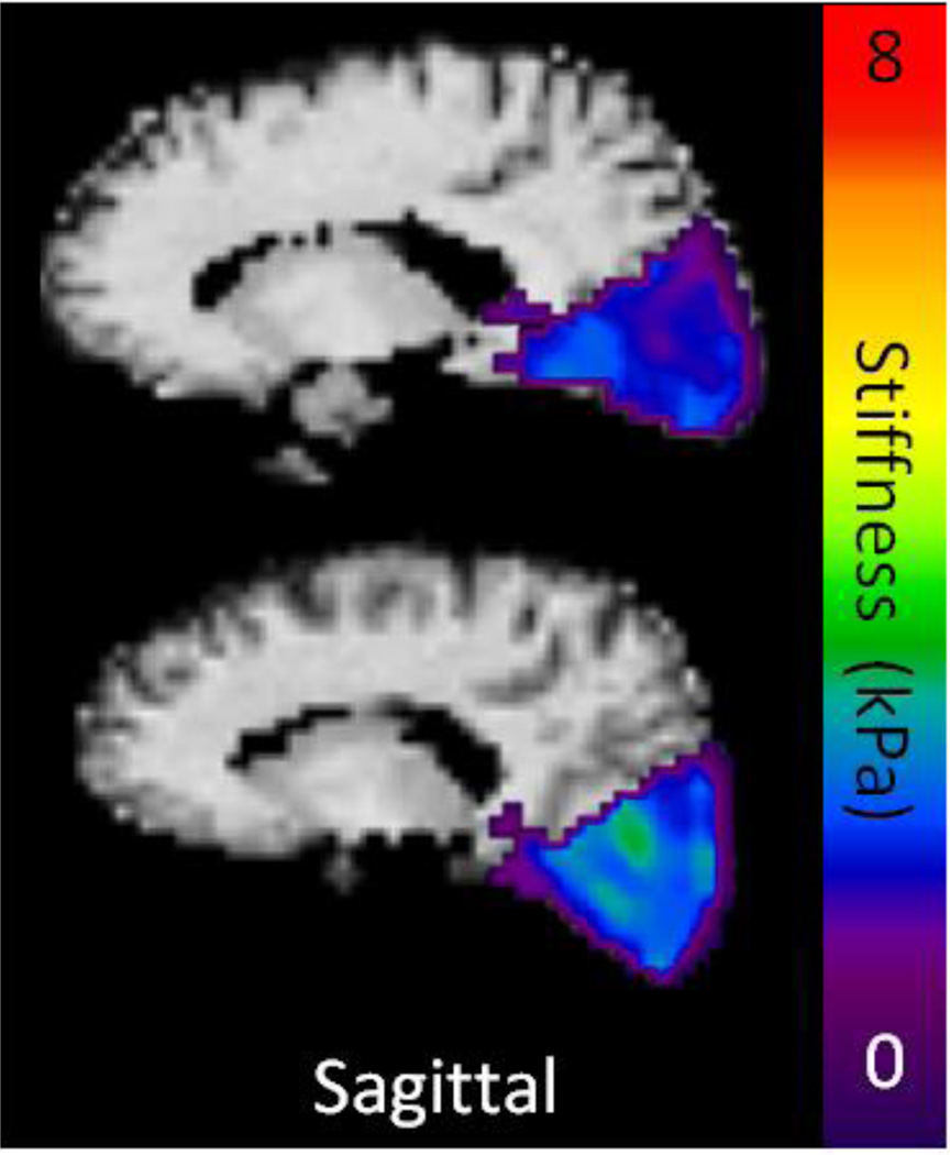
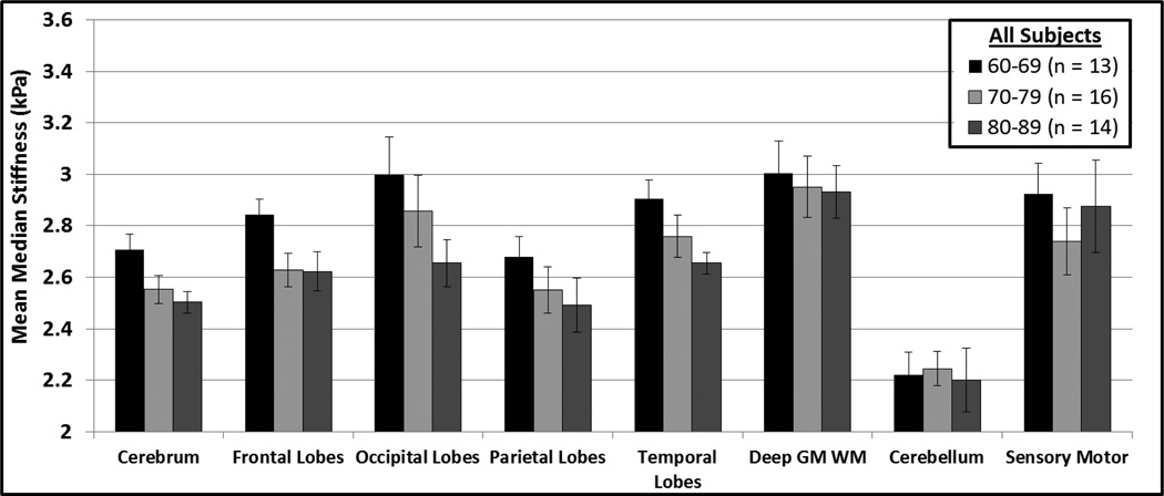
Changes in tissue composition and cellular architecture have been associated with neurological disease, and these in turn can affect biomechanical properties. Natural biological factors such as aging and an individual's sex also affect underlying tissue biomechanics in different brain regions. Understanding the normal changes is necessary before determining the efficacy of stiffness imaging for neurological disease diagnosis and therapy monitoring. The objective of this study was to evaluate global and regional changes in brain stiffness as a function of age and sex, using improved MRE acquisition and processing that have been shown to provide median stiffness values that are typically reproducible to within 1% in global measurements and within 2% for regional measurements. Furthermore, this is the first study to report the effects of age and sex over the entire cerebrum volume and over the full frontal, occipital, parietal, temporal, deep gray matter/white matter (insula, deep gray nuclei and white matter tracts), and cerebellum volumes. In 45 volunteers, we observed a significant linear correlation between age and brain stiffness in the cerebrum (P<.0001), frontal lobes (P<.0001), occipital lobes (P=.0005), parietal lobes (P=.0002), and the temporal lobes (P<.0001) of the brain. No significant linear correlation between brain stiffness and age was observed in the cerebellum (P=.74), and the sensory-motor regions (P=.32) of the brain, and a weak linear trend was observed in the deep gray matter/white matter (P=.075). A multiple linear regression model predicted an annual decline of 0.011 ± 0.002 kPa in cerebrum stiffness with a theoretical median age value (76 years old) of 2.56 ± 0.08 kPa. Sexual dimorphism was observed in the temporal (P=.03) and occipital (P=.001) lobes of the brain, but no significant difference was observed in any of the other brain regions (P>.20 for all other regions). The model predicted female occipital and temporal lobes to be 0.23 kPa and 0.09 kPa stiffer than males of the same age, respectively. This study confirms that as the brain ages, there is softening; however, the changes are dependent on region. In addition, stiffness effects due to sex exist in the occipital and temporal lobes.
Keywords: Aging; Brain MRE; Brain stiffness; Elastography; Gender bias; Sexual dimorphism.
Copyright © 2015 The Authors. Published by Elsevier Inc. All rights reserved.
Figures
References
-
- Allen JS, Bruss J, Brown CK, Damasio H. Normal neuroanatomical variation due to age: The major lobes and a parcellation of the temporal region. Neurobiol Aging. 2005;26:1245–1260. - PubMed
-
- Allen JS, Damasio H, Grabowski TJ, Bruss J, Zhang W. Sexual dimorphism and asymmetries in the gray-white composition of the human cerebrum. Neuroimage. 2003;18:880–894. - PubMed
-
- Ashburner J, Friston KJ. Unified segmentation. Neuroimage. 2005;26:839–851. - PubMed
-
- Bao AM, Swaab DF. Sex differences in the brain, behavior, and neuropsychiatric disorders. Neuroscientist. 2010;16:550–565. - PubMed
Publication types
MeSH terms
Grants and funding
LinkOut - more resources
Full Text Sources
Other Literature Sources
Medical
