

Combining endometrium sampling device and SurePath preparation to screen for endometrial carcinoma: a validation study
- PMID: 25698198
- PMCID: PMC4834777
- DOI: 10.4103/0366-6999.151664
Combining endometrium sampling device and SurePath preparation to screen for endometrial carcinoma: a validation study
Abstract
Background: The aim of this study was to compare specimen adequacy of SAP-1 provided for cytology with that of dilation and curettage (D & C) or hysteroscopy for histology, and evaluate the accuracy of combining endometrium sampling by SAP-1 and liquid-based cytology using SurePath preparation for screening endometrial carcinoma and its precursor.
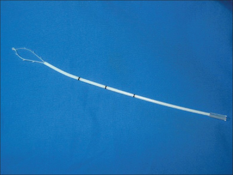
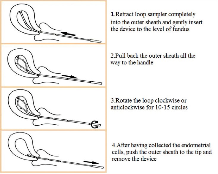
Methods: Endometrial specimens from women (n = 1514) with risk factors were obtained using an SAP-1 device for cytological analysis; histological samples were obtained from 375 of these women who underwent D & C or hysteroscopy. Cytological specimens were prepared to liquid-based smear using SurePath technology and stained by Papanicolaou. Histological samples were processed in routine pathology and stained by hematoxylin and eosin.
Results: Adequate specimens for cytology were obtained from 1458/1541 patients (96.3%), while adequate samples for pathology were obtained from 285/375 patients (76%). However, for postmenopausal women, 1006 of 1045 cytology (86.3%) were adequate, 153 of 238 histology (64.3%) were adequate, it was easier to collect cytological specimens than histological specimens (P < 0.05). The accuracy of endometrial cytology for detecting endometrial carcinoma and its precursor was 92.4% (sensitivity, 73%; specificity, 95.8%; positive predictive value, 75%; and negative predictive value, 95.3%).
Conclusions: Endometrial cytology using SAP-1 sampling and SurePath preparation may be a reliable approach for screening patients with endometrial carcinoma and its precursor.
Conflict of interest statement
Figures
References
-
- Siegel R, Ma J, Zou Z, Jemal A. Cancer statistics, 2014. CA Cancer J Clin. 2014;64:9–29. - PubMed
-
- Committee on Practice Bulletins – Gynecology. ACOG practice bulletin number 131: Screening for cervical cancer. Obstet Gynecol. 2012;120:1222–38. - PubMed
-
- Kurman RJ, McConnell TG. Precursors of endometrial and ovarian carcinoma. Virchows Arch. 2010;456:1–12. - PubMed
-
- Jarboe EA, Mutter GL. Endometrial intraepithelial neoplasia. Semin Diagn Pathol. 2010;27:215–25. - PubMed
-
- Zhao J. The diagnostic system of endometrial cytology. China J Reprod Health. 2006;17:6–8.
MeSH terms
LinkOut - more resources
Full Text Sources
Other Literature Sources
Medical
Miscellaneous
