. 2015 Apr:66:26-42.
doi: 10.1016/j.peptides.2015.01.009. Epub 2015 Feb 16.

A structure-function study of PACAP using conformationally restricted analogs: Identification of PAC1 receptor-selective PACAP agonists

Affiliations

A structure-function study of PACAP using conformationally restricted analogs: Identification of PAC1 receptor-selective PACAP agonists

Irene Ramos-Álvarez et al. Peptides. 2015 Apr.

Abstract

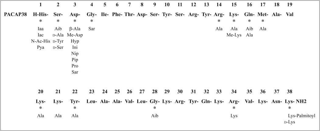
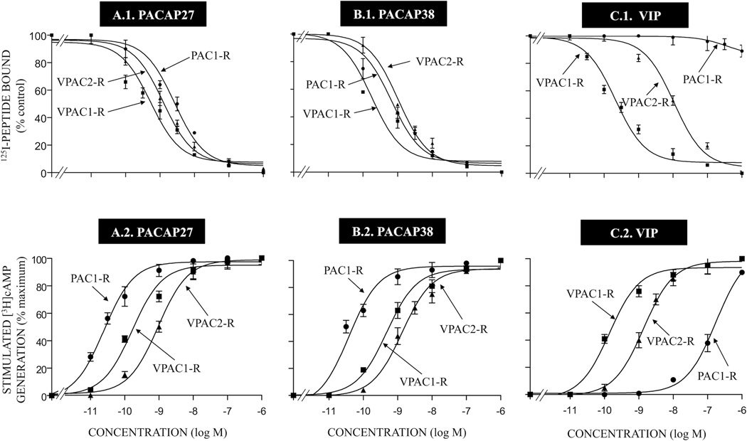
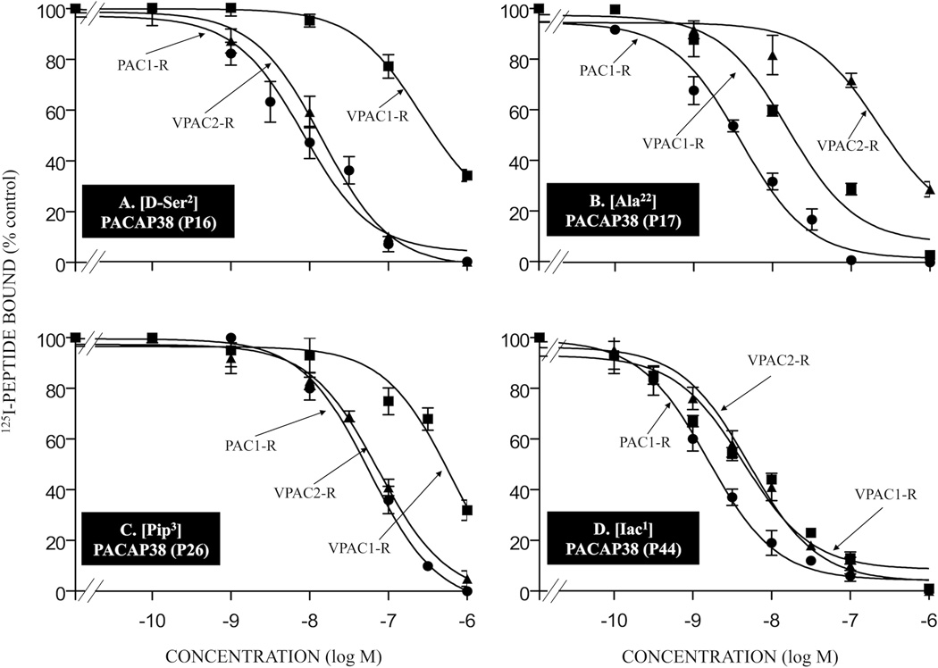
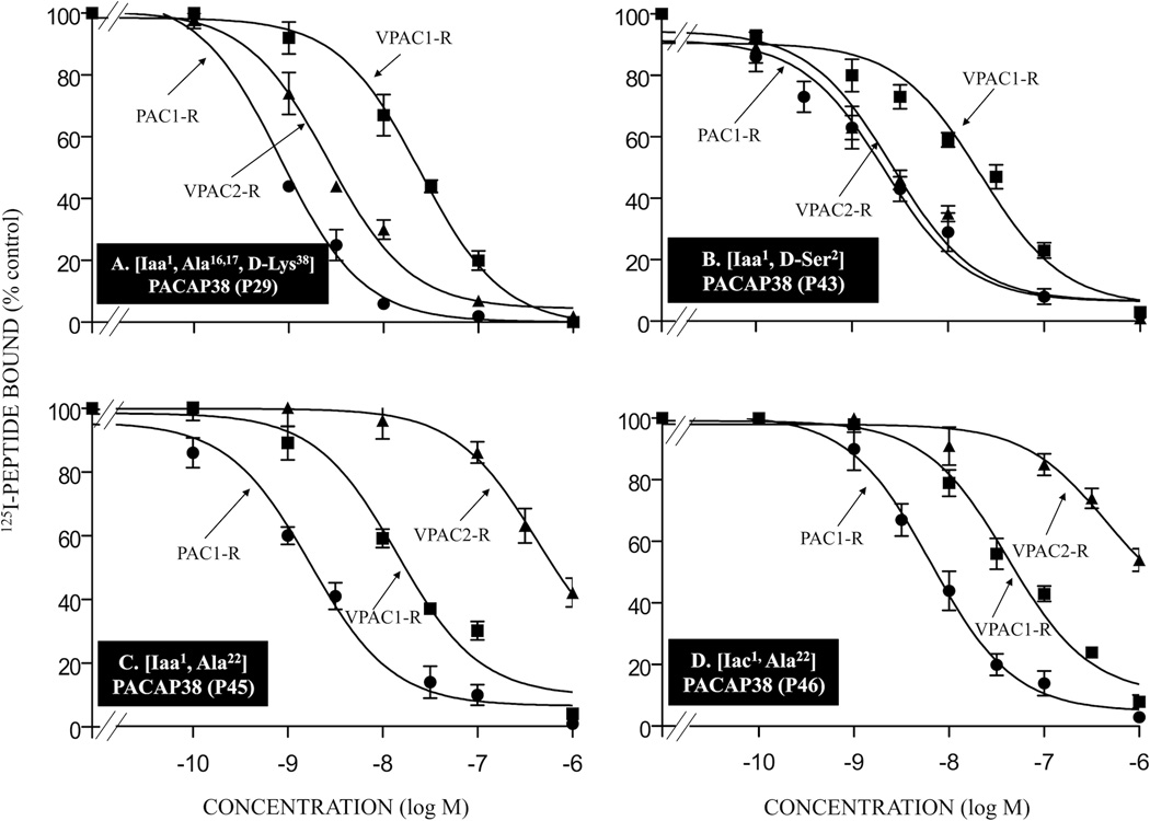
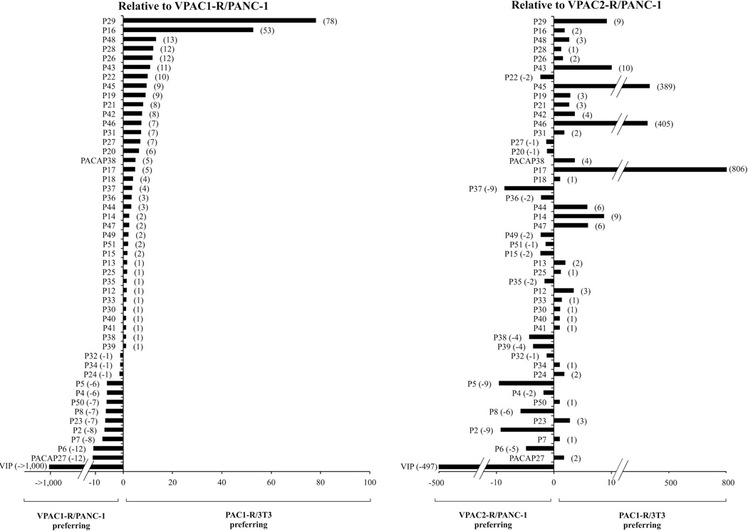
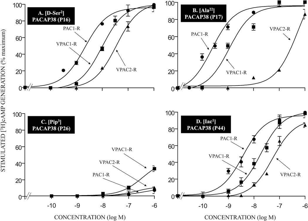
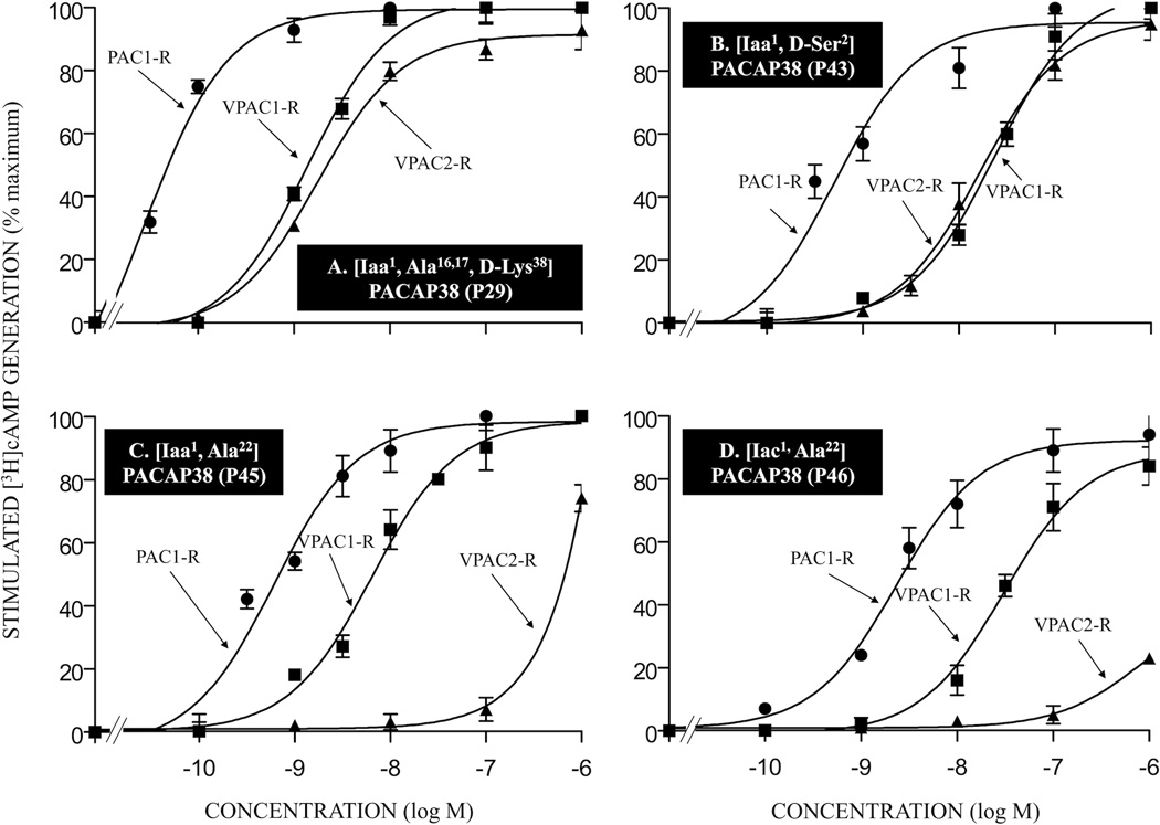
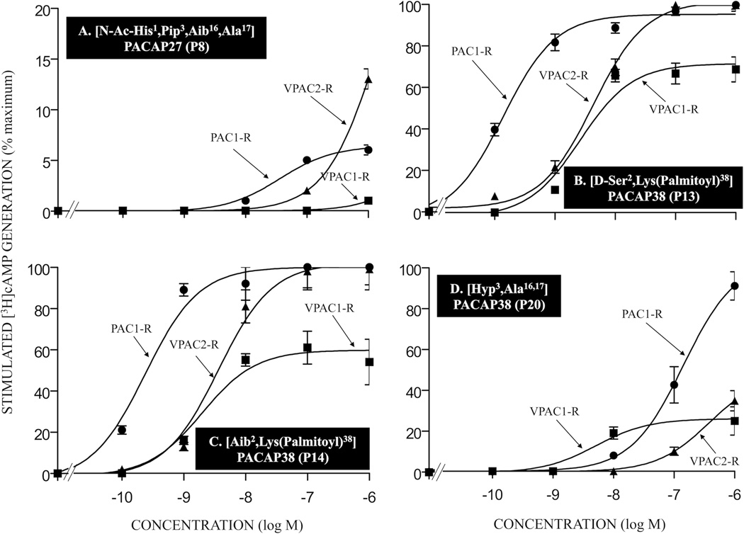
Pituitary adenylate cyclase-activating polypeptide (PACAP) has widespread physiological/pathophysiological actions and there is increased interest for its use therapeutically, especially in the CNS (neuroprotection). Unfortunately, no selective PACAP-analogs exist for PACAP-preferring PAC1-receptors, primarily because of its high sequence identity to VIP and particularly, because of the inability of structure-function studies to separate the pharmacophore of PAC1-R from VPAC1-R, which has high affinity for PACAP and VIP. The present study attempted to develop PAC1-R-selective agonists primarily by making conformationally restricted PACAP-analogs in positions important for receptor-selectivity/affinity. Forty-six PACAP-related-analogs were synthesized with substitutions in positions 1-4, 14-17, 20-22, 28, 34, 38 and receptor-selectivity determined in PAC1-R,VPAC1-R,VPAC2-R-transfected or native cells from binding or cAMP-generation experiments. Fifteen PACAP-analogs had 6-78-fold higher affinities for PAC1-R than VPAC1-R and 13 were agonists. Although binding-affinities correlated significantly with agonist potency, the degree of receptor-spareness varied markedly for the different PACAP-analogs, resulting in selective potencies for activating the PAC1 receptor over the VPAC1 receptor from 0- to 103-fold. In addition, a number of PACAP-analogs were identified that had high selectivity for PAC1-R over VPAC2-R as well as PACAP-analogs that could prove more useful therapeutically because of substitutions known to extend their half-lives (substitutions at potential sites of proteolysis and attachment of long-chain fatty acids). This study provides for the first time a separation of the pharmacophores for PAC1-R and VPAC1-R, resulting in PACAP-related analogs that are PAC1-R-preferring. Some of these analogs, or their modifications, could prove useful as therapeutic agents for various diseases.

Keywords: Neuroprotection; PACAP; Stroke; Structure–function study; Traumatic brain injury; Vasoactive intestinal peptide.


Figures

Figure 1
Figure 1
Positions of amino acid substitutions in PACAP38. The positions of substitutions made either alone or in combination. Abbreviations used: Ac, acetyl; Aib, α-amino-isobutyric acid (α-methylalanine); Ala, alanine; Arg, arginine; Asn, asparagine; Asp, aspartic acid; Gln, glutamine; Gly, glycine; His, histidine; Hyp, hydroxyproline; Iaa, 4-imidazole acetic acid; Iac, 4-imidazole acrylic acid; Ini, isonipecotic acid (piperidine-4-carboxylic acid); Ile, isoleucine; Leu, leucine; Lys, lysine; Me, methyl; Met, methionine; Nip, nipecotic acid (piperidine-3-carboxylic acid); PACAP, pituitary adenylate cyclase-activating polypeptide; Phe, phenylalanine; Pip, pipecolic acid (piperidine-2-carboxylic acid); Pro, proline; Pya, 3-pyridyl acetic acid; Sar, sarcosine (N-methylglycine); Ser, serine; Thr, threonine; Tyr, tyrosine; Val, valine.
Figure 2
Figure 2
Comparison of the ability of PACAP27, PACAP38 and VIP to inhibit binding of 125I-labeled peptide [upper panels] and their ability to stimulate [3H]cAMP accumulation [lower panels] in cells containing PAC1, VPAC1, VPAC2 receptors. Receptor binding was performed with 50 pM 125I-ligand as described in Materials and Methods for PAC1-R cells, VPAC1-R/PANC-1 cells and VPAC2-R/PANC-1 cells. Results are expressed as the percentage of saturable binding without unlabeled peptide added (percent control). Stimulation of cAMP accumulation [lower panels] was performed as described in Materials and Methods using 2 µCi/ml [3H]adenine. Values are expressed as percentage of maximal stimulated [3H]cAMP accumulation by 1 µM PACAP38. Control and 1 µM PACAP38 (as maximum) stimulated values for PAC1-R cells were 5328 ± 828 and 21717 ± 8759 dpm, respectively; for VPAC1-R/PANC-1 cells, 2020 ± 364 and 10084 ± 5213, respectively and for VPAC2-R/PANC-1 cells, 4444 ± 814 and 28951 ± 5213 dpm, respectively. The results are the mean and S.E.M. from at least three separate experiments. Abbreviations: see legends for Fig. 1 and Table 1.
Figure 3
Figure 3
Affinities of various single amino acid substituted PACAP-related analogs for PAC1-R, VPAC1-R and VPAC2-R. Briefly, cells containing PAC1-R, VPAC1-R and VPAC2-R were incubated for 60 min at room temperature with 50 pM 125I-ligand, with or without the indicated concentrations of unlabeled single amino acid substituted PACAP38 analogs. The results for the PACAP-analogs P16, P17, P26, and P44 are shown in Table 1. The experimental conditions and expression of the results are the same as in the legend for Fig. 2. Abbreviations: see legend for Table 1.
Figure 4
Figure 4
Affinities of various double- or multi-substituted PACAP-related analogs for PAC1-R, VPAC1-R and VPAC2-R. The experimental conditions were similar to those in the legend for Fig. 2 and Fig. 3. The values are the mean and S.E.M. from at least three separate experiments. The results for PACAP-analogs P29, P43, P45, and P46 are shown in Tables 1 and 2. Abbreviations: see legends for Fig. 1 and Table 1.
Figure 5
Figure 5
Relative affinities of PACAP-related analogs for VPAC1-R/PANC-1 or VPAC2-R/PANC-1 cells compared to affinity for PAC1-R cells. The results are expressed as the ratio of the affinity (IC50) for VPAC1-R/PANC-1(Left panel) or VPAC2-R /PANC-1(Right panel) cells divided by the affinity (IC50) for PAC1-R cells. The results are ordered by selectivity for PAC1-R cells compared to VPAC1-R PANC-1.
Figure 6
Figure 6
The potency of various single substituted PACAP-analogs for PAC1-R, VPAC1-R and VPAC2-R. Briefly, stably transfected PAC1-R, VPAC1-R/PANC-1 or VPAC2-R/PANC-1 cells were preincubated for 48 hours with [3H]adenine, washed and incubated for 60 minutes at 37°C with or without the indicated concentrations of the various PACAP38 analogs. The experimental conditions and expression of results were as describe in the legend Fig. 2. Values are means and S.E.M. from at least three separate experiments. The results for PACAP-related analogs P16, P17, P26, and P44 are shown in Table 3. Abbreviations: see legends for Fig. 1 and Table 1.
Figure 7
Figure 7
The potency of various double- or multi-substituted PACAP-analogs for PAC1-R, VPAC1-R and VPAC2-R. The experimental conditions were similar to those in the legends for Fig. 2 and Fig. 6. The values are the mean and S.E.M. from at least three separate experiments. The results for PACAP-analogs P29, P43, P45, and P46 are shown in Tables 2 and 3. Abbreviations: see legends for Fig. 1, Table 1 and Table 2.
Figure 8
Figure 8
The potency of various double- or multi-substituted PACAP-analogs for PAC1-R, VPAC1-R and VPAC2-R. The experimental conditions were similar to those in the legend to Fig. 2 and Fig. 6. Values are the mean and S.E.M. from at least three separate experiments. The results for PACAP-analogs P8, P13, P14, and P20 are shown in Tables 1 and 2. Abbreviations: see legends for Fig. 1, Table 1 and Table 2.
Figure 9
Figure 9
Comparison of the ability of PACAP38 and various single- or double-substituted PACAP-analogs to stimulate [3H]IP generation and cAMP generation in PAC1-R cells. For stimulation of cAMP, the cells were incubated with each ligand at the indicated concentration for 60 minutes. cAMP generation was determined in the indicated cell type loaded with [3H]adenine for 48 hours as described under Materials and Methods. [3H]IP was determined after a 60-minute incubation at 37°C after loading the cells for 48 hours with 3 mCi/ml myo-[2-3H]inositol as described in Materials and Methods. The control and maximal stimulated [3H]IP values for PAC1-R was 4047 ± 261 and 145873 ± 9962, respectively. The control and maximal cAMP generation values for PAC1-R was 5328 ± 828 and 21717 ± 8759, respectively. The values are the mean and S.E.M. from at least three separate experiments. The results are shown for PACAP38 and PACAP-analogs P42, P43 and P45. Abbreviations: see legends for Fig. 1, Table 1 and Table 2.

References

    1. Ahren B. Role of pituitary adenylate cyclase-activating polypeptide in the pancreatic endocrine system. Ann N Y Acad Sci. 2008;1144:28–35. - PubMed
    1. Alvarez R, Daniels DV. A separation method for the assay of adenylylcyclase, intracellular cyclic AMP, and cyclic-AMP phosphodiesterase using tritium-labeled substrates. Anal Biochem. 1992;203:76–82. - PubMed
    1. Banki E, Degrell P, Kiss P, Kovacs K, Kemeny A, Csanaky K, et al. Effect of PACAP treatment on kidney morphology and cytokine expression in rat diabetic nephropathy. Peptides. 2013;42:125–130. - PubMed
    1. Banks WA, Kastin AJ, Komaki G, Arimura A. Passage of pituitary adenylate cyclase activating polypeptide1-27 and pituitary adenylate cyclase activating polypeptide1-38 across the blood-brain barrier. J Pharmacol Exp Ther. 1993;267:690–696. - PubMed
    1. Baun M, Pedersen MH, Olesen J, Jansen-Olesen I. Dural mast cell degranulation is a putative mechanism for headache induced by PACAP-38. Cephalalgia. 2012;32:337–345. - PubMed

Publication types

MeSH terms

Substances

LinkOut - more resources