Review
. 2015 Feb:10:47-55.
doi: 10.1016/j.coviro.2014.12.003. Epub 2015 Feb 17.

Origin, evolution and innate immune control of simian foamy viruses in humans

Affiliations
Review

Origin, evolution and innate immune control of simian foamy viruses in humans

Rejane Rua et al. Curr Opin Virol. 2015 Feb.

Abstract

Most viral pathogens that have emerged in humans have originated from various animal species. Emergence is a multistep process involving an initial spill-over of the infectious agent into single individuals and its subsequent dissemination into the human population. Similar to simian immunodeficiency viruses and simian T lymphotropic viruses, simian foamy viruses (SFV) are retroviruses that are widespread among non-human primates and can be transmitted to humans, giving rise to a persistent infection, which seems to be controlled in the case of SFV. In this review, we present current data on the discovery, cross-species transmission, and molecular evolution of SFV in human populations initially infected and thus at risk for zoonotic emergence.


Figures

None
Graphical abstract
Figure 1
Figure 1
Schematic representation of SFV genomic organization (chimpanzee strain). The SFV genome is flanked by two long terminal repeat (LTRs) which contain the unique 3′ (U3), repeated (R) and unique 5′ (U5) regions. gag encodes the full-length gag protein (74 kDa) and the shorter p70 protein. pol encodes the protease (PR)-reverse transcriptase (RT)-Rnase H protein and the integrase (INT). env encodes the leader peptide (LP), the surface glycoprotein (SU) and the transmembrane protein (TM). Two additional genes tas and bet encode proteins having regulatory functions. The transactivator Tas binds to the 5’LTR which activates the transcription of the structural genes gag, pol and env.
Figure 2
Figure 2
Mechanisms of action of antiviral factors against FV. (A) SFV life cycle starts with infection of a new cell by SFV particles containing RNA and/or DNA, decapsidation of the viral genome, reverse transcription of RNA genomes and integration of viral DNA into cellular DNA. Cellular activation of the internal promoter induces tas and bet transcription. The transactivator Tas then activates a second promoter located in the long-terminal repeat, which induces the synthesis of the Gag, Pol and Env structural proteins. After assembling, viral particles bud essentially from the endoplasmic reticulum, with a late reverse-transcription step giving rise to both DNA and RNA particles. (B) SFV is detected by plasmacytoid dendritic cells (not exclusively) which triggers IFN-I production. Several antiviral factors that can be induced by IFN-I are active against SFV. TRIM5α are antiviral proteins that prevent SFV decapsidation [50]. Apolipoprotein B-editing catalytic polypeptide-like subunit (APOBEC) enzymes act on the negative strand DNA produced by the viral reverse transcriptase, resulting in G-to-A mutations in the viral genome, with potential deleterious consequences. FV Bet protein has been shown to partially prevent APOBEC action [40, 41, 42, 43]. N-Myc interactor (NMI) as well as a member of the interferon-induced protein (IFP) family, IFP35, can inhibit the replication of the prototype and the bovine FV respectively, by direct interaction with the viral transactivator Tas [46, 47]. Tetherin is known to block many different types of enveloped viruses by tethering the budding virus at the cell membrane and is also efficient to prevent FV release [48, 49].
Figure 3
Figure 3
Observation of SFV and SFV-infected cells. BHK-21 cells were infected with a chimpanzee SFV strain isolated from an SFV-infected hunter [31]. A large syncytium with a ‘foamy’ aspect is visible using light microscopy (A). An immunofluorescent staining was performed as previously described [62]. SFV antigens are revealed in the green channel and nuclei are stained with DAPI (B). U87MG cells were infected by SFV-1 and used for electron microscopy (C,D). Scale bars represent 10 μm (A), 3 μm (B) or 300 nm (C,D).

References

    1. Wolfe N.D., Dunavan C.P., Diamond J. Origins of major human infectious diseases. Nature. 2007;447:279–283. - PMC - PubMed
    1. Locatelli S., Peeters M. Cross-species transmission of simian retroviruses: how and why they could lead to the emergence of new diseases in the human population. AIDS. 2012;26:659–673. - PubMed
    1. Enders J.F., Peebles T.C. Propagation in tissue cultures of cytopathogenic agents from patients with measles. Proc Soc Exp Biol Med. 1954;86:277–286. - PubMed
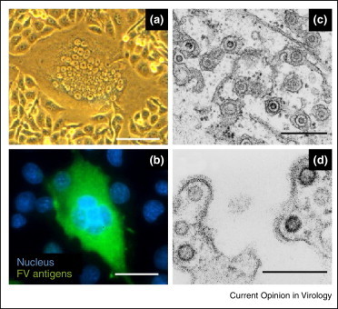
    1. Murray S.M., Picker L.J., Axthelm M.K., Hudkins K., Alpers C.E., Linial M.L. Replication in a superficial epithelial cell niche explains the lack of pathogenicity of primate foamy virus infections. J Virol. 2008;82:5981–5985. - PMC - PubMed
    2. This study provides convincing evidence of SFV replication in the oral cavity of SFV-infected animals. This is a major argument in favor of the special mode of transmission of this retrovirus, mainly by bites, as supported by epidemiological data.

    1. Choudhary A., Galvin T.A., Williams D.K., Beren J., Bryant M.A., Khan A.S. Influence of naturally occurring simian foamy viruses (SFVs) on SIV disease progression in the rhesus macaque (Macaca mulatta) model. Viruses. 2013;5:1414–1430. - PMC - PubMed

Publication types

MeSH terms