. 2015 Sep;9(3):217-31.
doi: 10.1007/s12079-015-0268-8. Epub 2015 Feb 20.

The MAPK and PI3K pathways mediate CNTF-induced neuronal survival and process outgrowth in hypothalamic organotypic cultures

Affiliations

The MAPK and PI3K pathways mediate CNTF-induced neuronal survival and process outgrowth in hypothalamic organotypic cultures

Jason M Askvig et al. J Cell Commun Signal. 2015 Sep.

Abstract

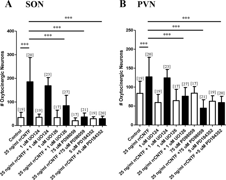
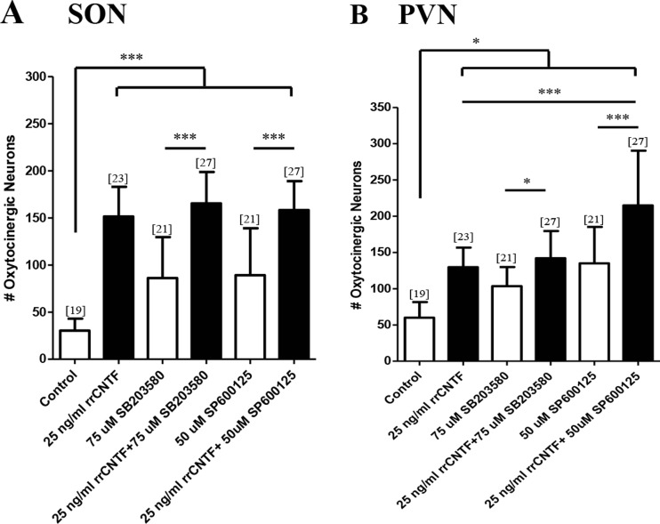
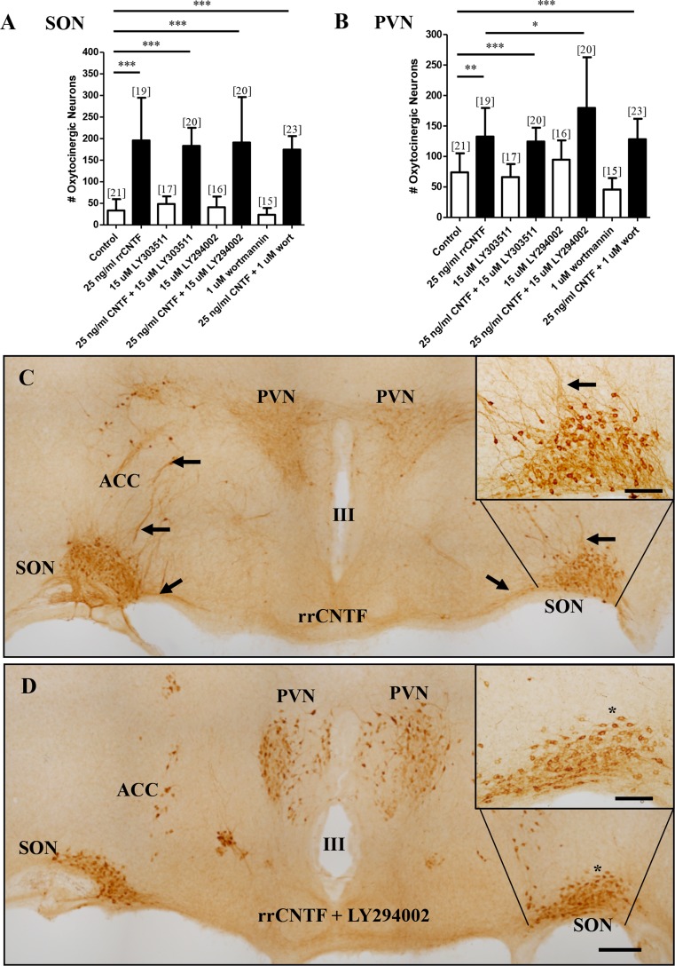
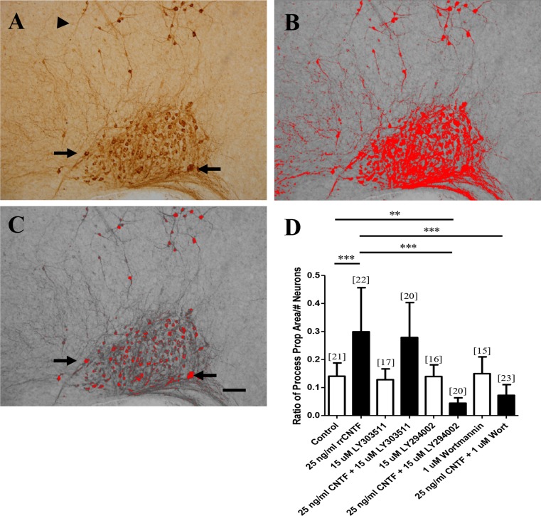
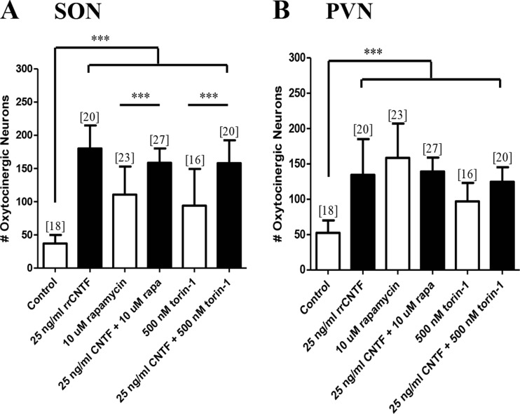
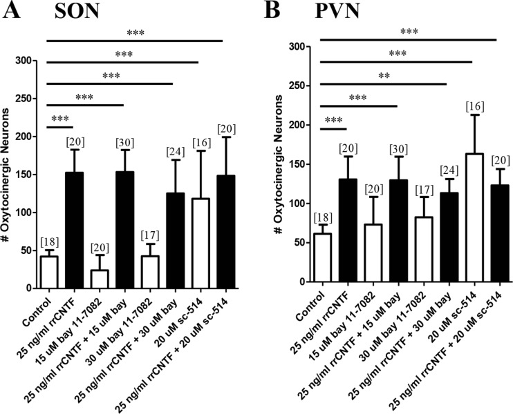
While collateral sprouting has been shown to occur in a variety of neuronal populations, the factor or factors responsible for mediating the sprouting response remain largely un-defined. There is evidence indicating that ciliary neurotrophic factor (CNTF) may play an important role in promoting neuronal survival and process outgrowth in neuronal phenotypes tested to date. We previously demonstrated that the astrocytic Jak-STAT pathway is necessary to mediate CNTF-induced oxytocinergic (OT) neuronal survival; however, the mechanism (s) of CNTF-mediated process outgrowth remain unknown. Our working hypothesis is that CNTF mediates differential neuroprotective responses via different intracellular signal transduction pathways. In order to test this hypothesis, we utilized stationary hypothalamic organotypic cultures to assess the contribution of the MAPK-ERK and PI3-AKT pathways to OT neuron survival and process outgrowth. Our results demonstrate that the MAPK-ERK½ pathway mediates CNTF-induced neuronal survival. Moreover, we show that inhibition of the p38-, JNK-MAPK, and mTOR pathways prevents loss OT neurons following axotomy. We also provide quantitative evidence indicating that CNTF promotes process outgrowth of OT neurons via the PI3K-AKT pathway. Together, these data indicate that distinct intracellular signaling pathways mediate diverse neuroprotective processes in response to CNTF.

Keywords: AKT; Axonal regeneration; CNTF; MAPK; NFkB; Neuronal survival; Organotypic culture; PI3K; STAT3.


Figures

Fig. 1
Fig. 1
The MAPK-ERK½ pathway is necessary to mediate the CNTF-induced survival of OT neurons. Immunohistochemical neuronal cell counts demonstrated that exogenous rrCNTF promoted the survival of OT neurons (SON: p < 0.0001; PVN: p < 0.0001) compared to control, while inhibition of the MAPK-ERK½ pathway with U0126 (SON: p < 0.0001; PVN: p < 0.0001), PD98059 (SON: p < 0.0001; PVN: p < 0.0001), and PD184352 (SON: p < 0.0001; PVN: p < 0.0001) significantly reduced the number of surviving OT neurons in the SON (a) and PVN (b) compared to the 25 ng/ml rrCNTF group. Column bars and error bars represent the mean and SD of [n] groups. PVN, paraventricular nucleus; SON, supraoptic nucleus. ***p < 0.0001
Fig. 2
Fig. 2
The p38- and JNK-MAPK pathways are not necessary to mediate the CNTF-induced survival of OT neurons. Immunohistochemical neuronal cell counts demonstrated that exogenous rrCNTF promoted the survival of OT neurons (SON: p < 0.0001; PVN: p < 0.05) compared to control, while inhibition of the p38- and JNK-MAPK pathways with SB203580 (SON: p < 0.0001; PVN: p < 0.0001) and SP600125 (SON: p < 0.0001; PVN: p < 0.0001), respectively, did not affect the number of surviving OT neurons in the SON (a) or PVN (b) compared to the 25 ng/ml rrCNTF group. However, when cultures were treated with the inhibitors alone, there was a statistically significant increase in the number of surviving OT neurons in the SON (A; SB203580: p < 0.0001; SP600125: p < 0.0001) and PVN (B; SB203580: p < 0.05; SP600125: p < 0.0001) compared to control. Column bars and error bars represent the mean and SD of [n] groups. PVN paraventricular nucleus, SON supraoptic nucleus. *p < 0.05, ***p < 0.0001
Fig. 3
Fig. 3
The PI3K-AKT pathway is not necessary to mediate the CNTF-induced survival of OT neurons. Immunohistochemical neuronal cell counts demonstrated that exogenous rrCNTF promoted the survival of OT neurons (SON: p < 0.0001; PVN: p < 0.01) compared to control, while inhibition of the PI3K-AKT pathway with LY294002 (SON: p = 0.6014; PVN: p = 0.4931) did not affect the number of surviving OT neurons in the SON (a) or PVN (b) compared to 25 ng/ml rrCNTF. In addition, the presence of wortmannin did not affect the number of surviving OT neurons in the SON (A; p = 0.8791); although there was a statistically significant increase in the number of surviving OT neurons in the PVN (B; p = 0.0373) compared to the 25 ng/ml rrCNTF group When visibly comparing the number of OT neurons between rrCNTF (c) and the rrCNTF plus LY294002 (d) groups, it is apparent that there are fewer neuronal processes present in the groups receiving PI3K inhibition compared to the rrCNTF group, even though they have the same number of surviving OT neurons (compare insets in C and D). Note that the representative images were obtained from approximately the same level of the magnocellular neurosecretory system, which is apparent when comparing the III ventricle between the images. Column bars and error bars represent the mean and SD of [n] groups. ACC accessory nuclei, PVN paraventricular nucleus, SON supraoptic nucleus. Scale bar C, D = 300 μm, inset = 100 μm. *p < 0.05, **p < 0.01, ***p < 0.0001
Fig. 4
Fig. 4
Inhibition of PI3K reduces the proportional area of OT-immunoreactive processes in the SON. a The image of the SON, captured such that the entire SON is centered in the bottom of the image. b A highlighted SON is shown following setting of the density. The density has been set to highlight the immunoreactive processes and somata, however, note that the analysis is a conservative estimate of the proportional area of neuronal processes because not all of the immunoreactive processes are highlighted (arrowheads, A, B). c A highlighted SON is shown following the setting of the density to highlight just the immunoreactive somata. Note that similar to their immunoreactive profiles, the nuclei are not highlighted by the density setting (arrows, A, C). d The ratio of proportional area of OT-immunoreactive processes to total number of neurons in the SON demonstrated that following administration of exogenous rrCNTF there was a significant increase of 112 % in the proportional area of OT-immunoreactive processes compared to control (p < 0.0001). The LY303511 molecule served as a control molecule for the LY294002 inhibitor and the proportional area of OT-immunoreactive processes in the LY303511 in conjunction with 25 ng/ml rrCNTF group did not differ from the 25 ng/ml rrCNTF group (p = 0.6496). However, when the PI3K inhibitors, LY294002 and wortmannin, were administered to the organotypic cultures in the presence of 25 ng/ml rrCNTF, there was a significant reduction in the proportional area of OT-immunoreactive processes in the SON compared to the 25 ng/ml rrCNTF group (p < 0.0001). Column bars and error bars represent the mean and SD of [n] groups. Scale bar = 100 μm. **p < 0.01, ***p < 0.0001
Fig. 5
Fig. 5
mTOR is not necessary to mediate CNTF-induced survival of OT neurons. Immunohistochemical neuronal cell counts demonstrated that exogenous rrCNTF promoted the survival of OT neurons (SON: p < 0.0001; PVN: p < 0.0001) compared to control, while inhibition of mTOR with rapamycin (SON: p = 0.1113; PVN: p = 0.6633) and torin-1 (SON: p = 0.1487; PVN: p = 0.4250) did not affect the number of surviving OT neurons in the SON (a) or PVN (b) compared to 25 ng/ml rrCNTF. However, when cultures were treated with the inhibitors alone, there was a significant increase in the number of surviving OT neurons in the SON (A; rapamycin: p < 0.0001; torin-1: p < 0.0001) and PVN (B; rapamycin: p < 0.0001; torin-1: p < 0.0001) compared to control. Column bars and error bars represent the mean and SD of [n] groups. PVN paraventricular nucleus, SON supraoptic nucleus. ***p < 0.0001
Fig. 6
Fig. 6
NF-κB is not necessary to mediate the CNTF-induced survival of OT neurons. Immunohistochemical neuronal cell counts demonstrated that exogenous rrCNTF promoted the survival of OT neurons (SON: p < 0.0001; PVN: p < 0.0001) compared to control, while inhibition of NF-κB with bay 11-7082 (15 μM: SON: p = 0.9228; PVN: p = 0.9003; 30 μM: SON: p = 0.0828; PVN: p = 0.0667) and sc-514 (SON: p = 0.7561; PVN: p = 0.3522) did not affect the number of surviving OT neurons in the SON (a) or PVN (b). However, when cultures were treated with sc-514 alone, there was a statistically significant increase in the number of surviving OT neurons in the SON (A; p < 0.0001) and PVN (B; p < 0.0001), compared to control. Column bars and error bars represent the mean and SD of [n] groups. PVN paraventricular nucleus, SON supraoptic nucleus. **p < 0.01, ***p < 0.0001

References

    1. Alonzi T, Middleton G, Wyatt S, Buchman V, Betz UA, Muller W, Musiani P, Poli V, Davies AM. Role of STAT3 and PI 3-kinase/Akt in mediating the survival actions of cytokines on sensory neurons. Mol Cell Neurosci. 2001;18:270–282. doi: 10.1006/mcne.2001.1018. - DOI - PubMed
    1. Armstrong SJ, Wiberg M, Terenghi G, Kingham PJ. Laminin activates NF-kappaB in Schwann cells to enhance neurite outgrowth. Neurosci Lett. 2008;439:42–46. doi: 10.1016/j.neulet.2008.04.091. - DOI - PubMed
    1. Askvig JM, Leiphon LJ, Watt JA. Neuronal activity and axonal sprouting differentially regulate CNTF and CNTF receptor complex in the rat supraoptic nucleus. Exp Neurol. 2012;233:243–252. doi: 10.1016/j.expneurol.2011.10.009. - DOI - PMC - PubMed
    1. Askvig JM, Lo DY, Sudbeck AW, Behm KE, Leiphon LJ, Watt JA. Inhibition of the Jak-STAT pathway prevents CNTF-mediated survival of axotomized oxytocinergic magnocellular neurons in organotypic cultures of the rat supraoptic nucleus. Exp Neurol. 2013;240:75–87. doi: 10.1016/j.expneurol.2012.10.023. - DOI - PMC - PubMed
    1. Asnaghi L, Calastretti A, Bevilacqua A, D’Agnano I, Gatti G, Canti G, Delia D, Capaccioli S, Nicolin A. Bcl-2 phosphorylation and apoptosis activated by damaged microtubules require mTOR and are regulated by Akt. Oncogene. 2004;23:5781–5791. doi: 10.1038/sj.onc.1207698. - DOI - PubMed