Influence of motivation on control hierarchy in the human frontal cortex
- PMID: 25698755
- PMCID: PMC6605591
- DOI: 10.1523/JNEUROSCI.2389-14.2015
Influence of motivation on control hierarchy in the human frontal cortex
Abstract
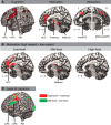
The frontal cortex mediates cognitive control and motivation to shape human behavior. It is generally observed that medial frontal areas are involved in motivational aspects of behavior, whereas lateral frontal regions are involved in cognitive control. Recent models of cognitive control suggest a rostro-caudal gradient in lateral frontal regions, such that progressively more rostral (anterior) regions process more complex aspects of cognitive control. How motivation influences such a control hierarchy is still under debate. Although some researchers argue that both systems work in parallel, others argue in favor of an interaction between motivation and cognitive control. In the latter case it is yet unclear how motivation would affect the different levels of the control hierarchy. This was investigated in the present functional MRI study applying different levels of cognitive control under different motivational states (low vs high reward anticipation). Three levels of cognitive control were tested by varying rule complexity: stimulus-response mapping (low-level), flexible task updating (mid-level), and sustained cue-task associations (high-level). We found an interaction between levels of cognitive control and motivation in medial and lateral frontal subregions. Specifically, flexible updating (mid-level of control) showed the strongest beneficial effect of reward and only this level exhibited functional coupling between dopamine-rich midbrain regions and the lateral frontal cortex. These findings suggest that motivation differentially affects the levels of a control hierarchy, influencing recruitment of frontal cortical control regions depending on specific task demands.
Keywords: cognitive control; control hierarchy; fMRI; lateral frontal cortex; motivation; reward.
Copyright © 2015 the authors 0270-6474/15/353207-11$15.00/0.
Figures


Similar articles
-
The Rostro-Caudal Axis of Frontal Cortex Is Sensitive to the Domain of Stimulus Information.Cereb Cortex. 2015 Jul;25(7):1815-26. doi: 10.1093/cercor/bht419. Epub 2014 Jan 22. Cereb Cortex. 2015. PMID: 24451658 Free PMC article.
-
Attentional control of task and response in lateral and medial frontal cortex: brain activity and reaction time distributions.Neuropsychologia. 2009 Aug;47(10):2089-99. doi: 10.1016/j.neuropsychologia.2009.03.019. Epub 2009 Apr 5. Neuropsychologia. 2009. PMID: 19467359
-
Transcranial magnetic stimulation reveals complex cognitive control representations in the rostral frontal cortex.Neuroscience. 2015 Aug 6;300:425-31. doi: 10.1016/j.neuroscience.2015.05.058. Epub 2015 May 30. Neuroscience. 2015. PMID: 26037799
-
Large-Scale Meta-Analysis of Human Medial Frontal Cortex Reveals Tripartite Functional Organization.J Neurosci. 2016 Jun 15;36(24):6553-62. doi: 10.1523/JNEUROSCI.4402-15.2016. J Neurosci. 2016. PMID: 27307242 Free PMC article. Review.
-
Cognitive control, hierarchy, and the rostro-caudal organization of the frontal lobes.Trends Cogn Sci. 2008 May;12(5):193-200. doi: 10.1016/j.tics.2008.02.004. Epub 2008 Apr 9. Trends Cogn Sci. 2008. PMID: 18403252 Review.
Cited by
-
The neural basis of motivational influences on cognitive control.Hum Brain Mapp. 2018 Dec;39(12):5097-5111. doi: 10.1002/hbm.24348. Epub 2018 Aug 18. Hum Brain Mapp. 2018. PMID: 30120846 Free PMC article.
-
Aversive motivation and cognitive control.Neurosci Biobehav Rev. 2022 Feb;133:104493. doi: 10.1016/j.neubiorev.2021.12.016. Epub 2021 Dec 12. Neurosci Biobehav Rev. 2022. PMID: 34910931 Free PMC article. Review.
-
Neural Pathways Linking Autonomous Exercise Motivation and Exercise-Induced Unhealthy Eating: A Resting-State fMRI Study.Brain Sci. 2024 Feb 27;14(3):221. doi: 10.3390/brainsci14030221. Brain Sci. 2024. PMID: 38539610 Free PMC article.
-
Integrative frontal-parietal dynamics supporting cognitive control.Elife. 2021 Mar 2;10:e57244. doi: 10.7554/eLife.57244. Elife. 2021. PMID: 33650966 Free PMC article.
-
Neural mechanisms underlying the reward-related enhancement of motivation when remembering episodic memories with high difficulty.Hum Brain Mapp. 2017 Jul;38(7):3428-3443. doi: 10.1002/hbm.23599. Epub 2017 Apr 4. Hum Brain Mapp. 2017. PMID: 28374960 Free PMC article.
References
Publication types
MeSH terms
Substances
Grants and funding
LinkOut - more resources
Full Text Sources
