

Brain amyloid-β burden is associated with disruption of intrinsic functional connectivity within the medial temporal lobe in cognitively normal elderly
- PMID: 25698758
- PMCID: PMC4331637
- DOI: 10.1523/JNEUROSCI.2092-14.2015
Brain amyloid-β burden is associated with disruption of intrinsic functional connectivity within the medial temporal lobe in cognitively normal elderly
Abstract
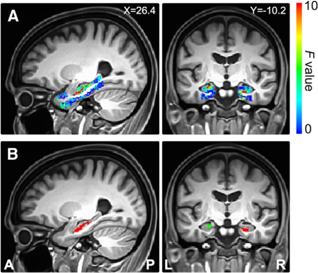
The medial temporal lobe is implicated as a key brain region involved in the pathogenesis of Alzheimer's disease (AD) and consequent memory loss. Tau tangle aggregation in this region may develop concurrently with cortical Aβ deposition in preclinical AD, but the pathological relationship between tau and Aβ remains unclear. We used task-free fMRI with a focus on the medical temporal lobe, together with Aβ PET imaging, in cognitively normal elderly human participants. We found that cortical Aβ load was related to disrupted intrinsic functional connectivity of the perirhinal cortex, which is typically the first brain region affected by tauopathies in AD. There was no concurrent association of cortical Aβ load with cognitive performance or brain atrophy. These findings suggest that dysfunction in the medial temporal lobe may represent a very early sign of preclinical AD and may predict future memory loss.
Keywords: Alzheimer's disease; amyloid; hippocampus; perirhinal cortex.
Copyright © 2015 the authors 0270-6474/15/353240-08$15.00/0.
Figures
Similar articles
-
Entorhinal Tau Pathology, Episodic Memory Decline, and Neurodegeneration in Aging.J Neurosci. 2018 Jan 17;38(3):530-543. doi: 10.1523/JNEUROSCI.2028-17.2017. Epub 2017 Nov 30. J Neurosci. 2018. PMID: 29192126 Free PMC article.
-
In vivo detection of microstructural correlates of brain pathology in preclinical and early Alzheimer Disease with magnetic resonance imaging.Neuroimage. 2017 Mar 1;148:296-304. doi: 10.1016/j.neuroimage.2016.12.026. Epub 2016 Dec 15. Neuroimage. 2017. PMID: 27989773 Free PMC article.
-
Frontotemporal network connectivity during memory encoding is increased with aging and disrupted by beta-amyloid.J Neurosci. 2013 Nov 20;33(47):18425-37. doi: 10.1523/JNEUROSCI.2775-13.2013. J Neurosci. 2013. PMID: 24259567 Free PMC article.
-
Alzheimer's disease.Subcell Biochem. 2012;65:329-52. doi: 10.1007/978-94-007-5416-4_14. Subcell Biochem. 2012. PMID: 23225010 Review.
-
MRI parcellation of ex vivo medial temporal lobe.Neuroimage. 2014 Jun;93 Pt 2:252-9. doi: 10.1016/j.neuroimage.2013.05.053. Epub 2013 May 21. Neuroimage. 2014. PMID: 23702414 Free PMC article. Review.
Cited by
-
Bridging Scales in Alzheimer's Disease: Biological Framework for Brain Simulation With The Virtual Brain.Front Neuroinform. 2021 Apr 1;15:630172. doi: 10.3389/fninf.2021.630172. eCollection 2021. Front Neuroinform. 2021. PMID: 33867964 Free PMC article. Review.
-
Mechanical property alterations across the cerebral cortex due to Alzheimer's disease.Brain Commun. 2020;2(1):fcz049. doi: 10.1093/braincomms/fcz049. Epub 2019 Dec 17. Brain Commun. 2020. PMID: 31998866 Free PMC article.
-
ABCA7 risk variant in healthy older African Americans is associated with a functionally isolated entorhinal cortex mediating deficient generalization of prior discrimination training.Hippocampus. 2019 Jun;29(6):527-538. doi: 10.1002/hipo.23042. Epub 2018 Dec 10. Hippocampus. 2019. PMID: 30318785 Free PMC article.
-
Sildenafil Improves Vascular and Metabolic Function in Patients with Alzheimer's Disease.J Alzheimers Dis. 2017;60(4):1351-1364. doi: 10.3233/JAD-161006. J Alzheimers Dis. 2017. PMID: 29036811 Free PMC article.
-
Positive Effect of Cognitive Reserve on Episodic Memory, Executive and Attentional Functions Taking Into Account Amyloid-Beta, Tau, and Apolipoprotein E Status.Front Aging Neurosci. 2021 May 28;13:666181. doi: 10.3389/fnagi.2021.666181. eCollection 2021. Front Aging Neurosci. 2021. PMID: 34122044 Free PMC article.
References
-
- Aizenstein HJ, Nebes RD, Saxton JA, Price JC, Mathis CA, Tsopelas ND, Ziolko SK, James JA, Snitz BE, Houck PR, Bi W, Cohen AD, Lopresti BJ, DeKosky ST, Halligan EM, Klunk WE. Frequent amyloid deposition without significant cognitive impairment among the elderly. Arch Neurol. 2008;65:1509–1517. doi: 10.1001/archneur.65.11.1509. - DOI - PMC - PubMed