

Alcohol decreases baseline brain glucose metabolism more in heavy drinkers than controls but has no effect on stimulation-induced metabolic increases
- PMID: 25698759
- PMCID: PMC4331638
- DOI: 10.1523/JNEUROSCI.4877-14.2015
Alcohol decreases baseline brain glucose metabolism more in heavy drinkers than controls but has no effect on stimulation-induced metabolic increases
Abstract
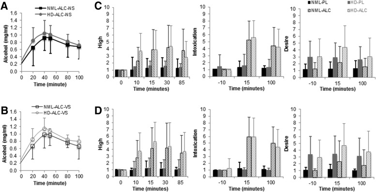
During alcohol intoxication, the human brain increases metabolism of acetate and decreases metabolism of glucose as energy substrate. Here we hypothesized that chronic heavy drinking facilitates this energy substrate shift both for baseline and stimulation conditions. To test this hypothesis, we compared the effects of alcohol intoxication (0.75 g/kg alcohol vs placebo) on brain glucose metabolism during video stimulation (VS) versus when given with no stimulation (NS), in 25 heavy drinkers (HDs) and 23 healthy controls, each of whom underwent four PET-(18)FDG scans. We showed that resting whole-brain glucose metabolism (placebo-NS) was lower in HD than controls (13%, p = 0.04); that alcohol (compared with placebo) decreased metabolism more in HD (20 ± 13%) than controls (9 ± 11%, p = 0.005) and in proportion to daily alcohol consumption (r = 0.36, p = 0.01) but found that alcohol did not reduce the metabolic increases in visual cortex from VS in either group. Instead, VS reduced alcohol-induced decreases in whole-brain glucose metabolism (10 ± 12%) compared with NS in both groups (15 ± 13%, p = 0.04), consistent with stimulation-related glucose metabolism enhancement. These findings corroborate our hypothesis that heavy alcohol consumption facilitates use of alternative energy substrates (i.e., acetate) for resting activity during intoxication, which might persist through early sobriety, but indicate that glucose is still favored as energy substrate during brain stimulation. Our findings are consistent with reduced reliance on glucose as the main energy substrate for resting brain metabolism during intoxication (presumably shifting to acetate or other ketones) and a priming of this shift in HDs, which might make them vulnerable to energy deficits during withdrawal.
Keywords: acetate metabolism; alcoholism; glial metabolism; glycolysis; positron emission tomography.
Copyright © 2015 the authors 0270-6474/15/353248-08$15.00/0.
Figures


Similar articles
-
Acute alcohol intoxication decreases glucose metabolism but increases acetate uptake in the human brain.Neuroimage. 2013 Jan 1;64:277-83. doi: 10.1016/j.neuroimage.2012.08.057. Epub 2012 Aug 28. Neuroimage. 2013. PMID: 22947541 Free PMC article.
-
Low doses of alcohol substantially decrease glucose metabolism in the human brain.Neuroimage. 2006 Jan 1;29(1):295-301. doi: 10.1016/j.neuroimage.2005.07.004. Epub 2005 Aug 8. Neuroimage. 2006. PMID: 16085426 Clinical Trial.
-
Regional brain metabolism during alcohol intoxication.Alcohol Clin Exp Res. 2000 Jun;24(6):822-9. Alcohol Clin Exp Res. 2000. PMID: 10888070 Clinical Trial.
-
Subjective response to alcohol challenge: a quantitative review.Alcohol Clin Exp Res. 2011 Oct;35(10):1759-70. doi: 10.1111/j.1530-0277.2011.01521.x. Epub 2011 Jul 20. Alcohol Clin Exp Res. 2011. PMID: 21777258 Free PMC article. Review.
-
Heart rate variability in alcohol use: A review.Pharmacol Biochem Behav. 2019 Jan;176:83-92. doi: 10.1016/j.pbb.2018.12.003. Epub 2018 Dec 6. Pharmacol Biochem Behav. 2019. PMID: 30529588 Review.
Cited by
-
Neuroimmunometabolism: A New Pathological Nexus Underlying Neurodegenerative Disorders.J Neurosci. 2022 Mar 9;42(10):1888-1907. doi: 10.1523/JNEUROSCI.0998-21.2022. Epub 2022 Jan 13. J Neurosci. 2022. PMID: 35027409 Free PMC article. Review.
-
Acute alcohol induces greater dose-dependent increase in the lateral cortical network functional connectivity in adult than adolescent rats.Addict Neurosci. 2023 Sep;7:100105. doi: 10.1016/j.addicn.2023.100105. Epub 2023 Jun 2. Addict Neurosci. 2023. PMID: 37576436 Free PMC article.
-
A critical review of ethanol effects on neuronal firing: A metabolic perspective.Alcohol Clin Exp Res (Hoboken). 2024 Mar;48(3):450-458. doi: 10.1111/acer.15266. Epub 2024 Jan 12. Alcohol Clin Exp Res (Hoboken). 2024. PMID: 38217065 Free PMC article. Review.
-
Paradoxical mTORC1-Dependent microRNA-mediated Translation Repression in the Nucleus Accumbens of Male Mice Consuming Alcohol Attenuates Glycolysis.Nat Commun. 2025 Jul 14;16(1):6116. doi: 10.1038/s41467-025-60337-9. Nat Commun. 2025. PMID: 40659615 Free PMC article.
-
Association Between Reduced Brain Glucose Metabolism and Cortical Thickness in Alcoholics: Evidence of Neurotoxicity.Int J Neuropsychopharmacol. 2019 Sep 1;22(9):548-559. doi: 10.1093/ijnp/pyz036. Int J Neuropsychopharmacol. 2019. PMID: 31369670 Free PMC article.
References
-
- Adams KM, Gilman S, Koeppe RA, Kluin KJ, Brunberg JA, Dede D, Berent S, Kroll PD. Neuropsychological deficits are correlated with frontal hypometabolism in positron emission tomography studies of older alcoholic patients. Alcohol Clin Exp Res. 1993;17:205–210. doi: 10.1111/j.1530-0277.1993.tb00750.x. - DOI - PubMed
Publication types
MeSH terms
Substances
Grants and funding
LinkOut - more resources
Full Text Sources
Miscellaneous