. 2015 Feb 4:15:15.
doi: 10.1186/s12935-015-0168-2. eCollection 2015.

Galangin, a novel dietary flavonoid, attenuates metastatic feature via PKC/ERK signaling pathway in TPA-treated liver cancer HepG2 cells

Affiliations

Galangin, a novel dietary flavonoid, attenuates metastatic feature via PKC/ERK signaling pathway in TPA-treated liver cancer HepG2 cells

Shang-Tao Chien et al. Cancer Cell Int. .

Abstract

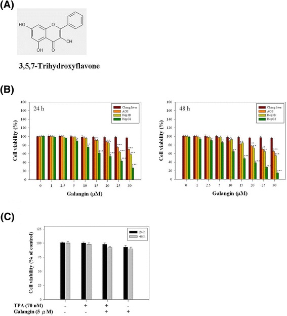
Background: Galangin (3,5,7-trihydroxyflavone) is a flavonoid compound found in high concentration in lesser galangal. The objective of this study was to investigate the ability of galangin to inhibit 12-O-tetradecanoylphorbol-13-acetate (TPA)-induced the invasion and metastasis of HepG2 liver cancer cells.

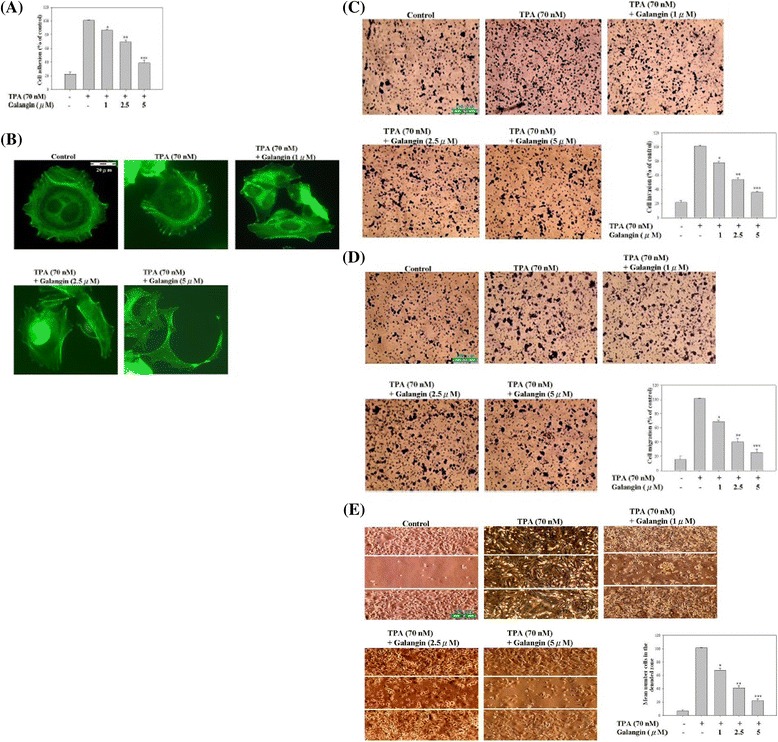
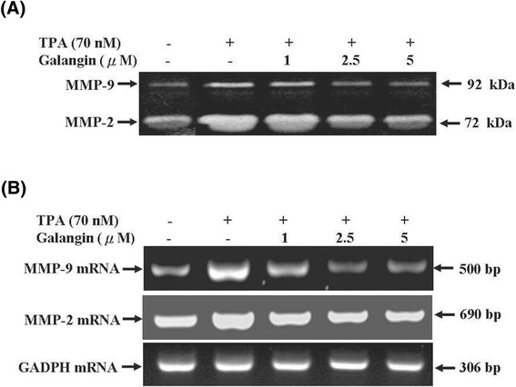
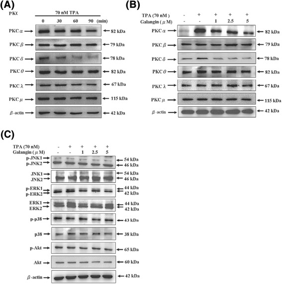
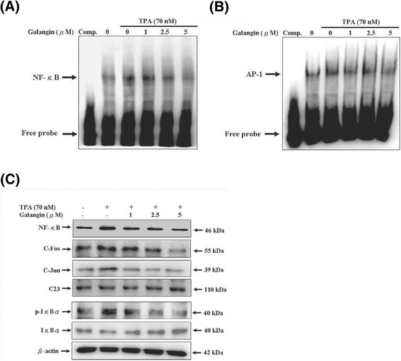
Results: First, using a cell-matrix adhesion assay, immunofluorescence assay, transwell-chamber invasion/migration assay, and wound healing assay, we observed that galangin exerted an inhibitory effect on TPA-induced cell adhesion, morphology/actin cytoskeleton arrangement, invasion and migration. Furthermore, the results of gelatin zymography and reverse transcriptase polymerase chain reaction (RT-PCR) assays showed that galangin reduced the TPA-induced enzyme activity of matrix metalloproteinase-2 (MMP-2) and matrix metalloproteinase-9 (MMP-9) in HepG2 cells; moreover, the messenger RNA level was downregulated. We also observed through a Western blotting assay that galangin strongly inhibited the TPA-induced protein expressions of protein kinase Cα (PKCα), protein kinase Cδ (PKCδ), phosphorylated extracellular signal-regulated kinase 1/2 (ERK1/2), the phospho-inhibitor of kappaBα (phospho-IκBα), c-Fos, c-Jun, and nuclear factor kappa B (NF-κB). Next, galangin dose-dependently inhibited the binding ability of NF-κB and activator protein 1 (AP-1) to MMP-2/MMP-9 promoters, respectively, resulting in the suppression of MMP-2/MMP-9 enzyme activity.

Conclusions: The results revealed that galangin effectively inhibited the TPA-induced invasion and migration of HepG2 cells through a protein kinase C/extracellular signal-regulated kinase (PKC/ERK) pathway. Thus, galangin may have widespread applications in clinical therapy as an anti-metastatic medicament.

Keywords: ERK; Galangin; Invasion; MMP-2; MMP-9; Migration; PKC-α; TPA.


Figures

Figure 1
Figure 1
Effect of galangin on the viability in four cell lines, Chang liver, AGS, Hep3B, and HepG2 cells. (A) Chemical structure of galangin. (B) Four cell lines were treated with or without various concentrations of galangin (0, 1, 2.5, 5, 10, 15, 20, 25, and 30 μM) for 24 and 48 h, separately. (C) HepG2 cells were treated with or without of drugs (70 nM TPA and 5 μM galangin) for 24 and 48 h. Thereafter, cell viability was determined by MTT assay. The survival cell number was directly proportional to formazan, which was measured spectrophotometrically at 563 nm. Values represent mean ± SD of three independent experiments. *p < 0.05, **p < 0.01, ***p < 0.001 compared with the untreated control (dose 0).
Figure 2
Figure 2
Effect of galangin on TPA-induced cell-matrix adhesion, cell morphology/actin cytoskeleton arrangement, invasion, and migration in HepG2 cells. Cells were treated with 70 nM TPA for 12 h and incubated in various concentrations of galangin (0, 1, 2.5, and 5 μM) for 24 h, and then were analysed for (A) cell-matrix adhesion, (B) immunofluorescence, scale bar: 20 μm, (C) transwell-chamber invasion, scale bar: 100 μm, (D) transwell-chamber migration, scale bar: 100 μm, and (E) wound-healing, scale bar: 100 μm. The aforementioned methods were described in “Materials and methods” section. Values are expressed as mean ± SD of three independent experiments. *p < 0.05, **p < 0.01, ***p < 0.001 compared with the untreated control (dose 0).
Figure 3
Figure 3
Effect of galangin on TPA-induced activity and expression of MMP-2/MMP-9 in HepG2 cells. (A) Cells were treated with 70 nM TPA for 12 h in serum-free medium and then incubated in various concentrations of galangin (0, 1, 2.5, and 5 μM) for 24 h. The conditioned media were collected and MMP-2/MMP-9 activities were determined by gelatin zymography. (B) Cells were treated with 70 nM TPA for 12 h and incubated in various concentrations of galangin (0, 1, 2.5, and 5 μM) for 12 h. And then, the RNA samples were extracted and subjected to a semi-quantitative RT-PCR for MMP-2 and MMP-9 with GADPH being an internal control.
Figure 4
Figure 4
Effect of galangin on TPA-induced the PKCα , PKCδ activation and ERK phosphorylation in HepG2 cells. (A) Cells were treated with TPA (70 nM) for various times, the protein levels of PKCα, PKCβ, PKCδ, PKCθ, PKCλ, and PKCμ in the membrane fraction were analyzed. (B) Cells were treated with various concentrations (0, 1, 2.5, and 5 μM) of galangin in the presence or absence of TPA (70 nM) for 6 h, the protein levels of PKCα, PKCβ, PKCδ, PKCθ, PKCλ, and PKCμ in the membrane fraction was analysed. (C) Cells were treated with TPA (70 nM) for 2 h in various concentrations (0, 1, 2.5, and 5 μM) of galangin for 6 h. The JNK phosphorylation, JNK, ERK phosphorylation, ERK, p38 phosphorylation, p38, Akt phosphorylation, and Akt were analysed by Western blotting. β-Actin was used as a loading control. The relative density of phosphorylated forms of JNK, ERK, and p38 were normalized to total values of JNK, ERK, and p38, which were determined by densitometric analysis.
Figure 5
Figure 5
Effect of galangin on the TPA-induced the DNA-binding activity of NF-κ B and AP-1/expressions of NF-κ B, c-Fos, c-Jun/Iκ phosphorylation and degradation in HepG2 cells. Nuclear extracts were prepared from HepG2 cells that treated with various concentrations of galangin (0, 1, 2.5, and 5 μM) in the presence of TPA (70 nM) for 12 h, and then used to analyze (A) NF-κB and (B) AP-1 DNA-binding activity by EMSA, as described in “Materials and methods” section. Lane 1: nuclear extracts incubated with 100-fold excess unlabeled consensus oligonucleotide (comp.) to confirm the binding specificity. Lane 2 represents nuclear extract from HepG2 cells in the absence of TPA (negative control). (C) Nuclear or cytosolic extracts were subjected to SDS-PAGE followed by western blotting with specific antobodies (anti-NF-κB, anti-c-Fos, anti-c-Jun anti-p-IκBα, anti-IκBα). C23 and β-actin were used as internal control.

Similar articles

Cited by

References

    1. Song PM, Zhang Y, He YF, Bao HM, Luo JH, Liu YK, et al. Bioinformatics analysis of metastasis-related proteins in hepatocellular carcinoma. World J Gastroenterol. 2008;14:5816–22. doi: 10.3748/wjg.14.5816. - DOI - PMC - PubMed
    1. Tung-Ping Poon R, Fan ST, Wong J. Risk factors, prevention and management of postoperative recurrence after resection of hepatocellular carcinoma. Ann Surg. 2000;232:10–24. doi: 10.1097/00000658-200007000-00003. - DOI - PMC - PubMed
    1. Huang SC, Ho CT, Lin-Shiau SY, Lin JK. Carnosol inhibits the invasion of B16/F10 mouse melanoma cells by suppressing metalloproteinase-9 through down- regulating nuclear factor-kappa B and c-Jun. Biochem Pharmacol. 2005;69:221–32. doi: 10.1016/j.bcp.2004.09.019. - DOI - PubMed
    1. Maeta H, Ohgi S, Terada T. Protein expression of matrix metalloproteinases 2 and 9 and tissue inhibitors of metalloproteinase 1 and 2 in papillary thyroid carcinomas. Virchows Arch. 2001;438:121–8. doi: 10.1007/s004280000286. - DOI - PubMed
    1. Egeblad M, Werb Z. New functions for the matrix metalloproteinases in cancer progression. Nat Rev Cancer. 2002;2:161–74. doi: 10.1038/nrc745. - DOI - PubMed