. 2015 Feb 2:8:2.
doi: 10.3389/fnmol.2015.00002. eCollection 2015.

Highly efficient method for gene delivery into mouse dorsal root ganglia neurons

Affiliations

Highly efficient method for gene delivery into mouse dorsal root ganglia neurons

Lingli Yu et al. Front Mol Neurosci. .

Erratum in

Abstract

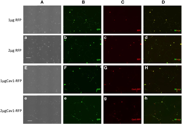
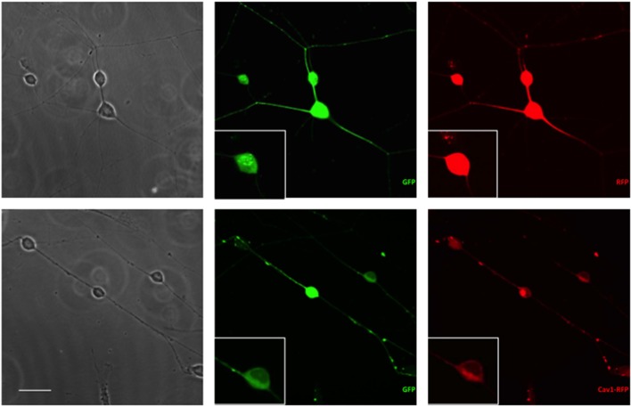
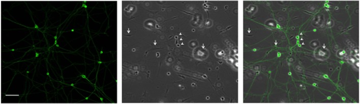
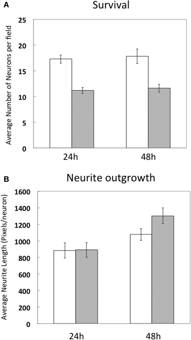
The development of gene transfection technologies has greatly advanced our understanding of life sciences. While use of viral vectors has clear efficacy, it requires specific expertise and biological containment conditions. Electroporation has become an effective and commonly used method for introducing DNA into neurons and in intact brain tissue. The present study describes the use of the Neon® electroporation system to transfect genes into dorsal root ganglia neurons isolated from embryonic mouse Day 13.5-16. This cell type has been particularly recalcitrant and refractory to physical or chemical methods for introduction of DNA. By optimizing the culture condition and parameters including voltage and duration for this specific electroporation system, high efficiency (60-80%) and low toxicity (>60% survival) were achieved with robust differentiation in response to Nerve growth factor (NGF). Moreover, 3-50 times fewer cells are needed (6 × 10(4)) compared with other traditional electroporation methods. This approach underlines the efficacy of this type of electroporation, particularly when only limited amount of cells can be obtained, and is expected to greatly facilitate the study of gene function in dorsal root ganglia neuron cultures.

Keywords: EGFP expression; Nerve growth factor (NGF); dorsal root ganglion (DRG) neuron; electroporation; gene expression; nucleofection; primary neurons; transfection.


Figures

Figure 1
Figure 1
Expression of fluorescent proteins in DRG neurons. Neon transfection leads to efficient electroporation of E14.5 DRG neurons. Neurons were transfected with 0.5 μg EGFP along with 1 or 2 μg of RFP or 1 or 2 μg Cav1-RFP. 24 h after electroporation, cells were fixed and observed under the microscope. Column A: Phase contrast image. Column B: GFP fluorescence. Column C: RFP fluorescence. Column D: Merge of GFP and RFP Fluorescence. Scale Bars represent 200 μm.
Figure 2
Figure 2
Expression of fluorescent proteins in DRG neurons. Neurons were transfected with 1 μg RFP or Cav1-RFP combined with 0.5 μg EGFP. 24 h after electroporation, cells were fixed and observed under the microscope. Scale Bar represents 50 μm.
Figure 3
Figure 3
Representative DRG neurons in culture. DRG neurons were dissected and maintained in culture. 24 h later, cells were fixed and labeled with anti-β III tubulin antibody. Arrows and arrowheads point, respectively, to glial and neuronal cells. Scale Bar represents 100 μm.
Figure 4
Figure 4
Survival and differentiation of DRG neurons after electroporation. DRG neurons were prepared and directly cultured without transfection (white bars) or transfected with 1 μg RFP combined with 0.5 μg EGFP (hashed bars). 24 and 48 h after electroporation, cells were fixed and neuron survival (A) and average neurite length per neuron (1pixel = 0.318 μm) (B) were calculated compared to naïve, non-transfected neurons. Results were obtained from three independent experiments. Data represent mean ± S.E.M. derived from 40 images representative of 692 non-transfected neurons (24 h) and 39 images representative of 439 GFP/RFP expressing neurons and 29 images representative of 523 non-transfected neurons and 31 images representative of 360 GFP/RFP expressing neurons of naïve, non-transfected controls (48 h).

Similar articles

Cited by

References

    1. Bertram B., Wiese S., von Holst A. (2012). High-efficiency transfection and survival rates of embryonic and adult mouse neural stem cells achieved by electroporation. J. Neurosci. Methods 209, 420–427. 10.1016/j.jneumeth.2012.06.024 - DOI - PubMed
    1. Boulaire J., Balani P., Wang S. (2009). Transcriptional targeting to brain cells: engineering cell type-specific promoter containing cassettes for enhanced transgene expression. Adv. Drug Deliv. Rev. 61, 589–602. 10.1016/j.addr.2009.02.007 - DOI - PubMed
    1. Butcher A. J., Torrecilla I., Young K. W., Kong K. C., Mistry S. C., Bottrill A. R., et al. . (2009). N-methyl-D-aspartate receptors mediate the phosphorylation and desensitization of muscarinic receptors in cerebellar granule neurons. J. Biol. Chem. 284, 17147–17156. 10.1074/jbc.M901031200 - DOI - PMC - PubMed
    1. Castellani V., Falk J., Rougon G. (2004). Semaphorin3A-induced receptor endocytosis during axon guidance responses is mediated by L1 CAM. Mol. Cell. Neurosci. 26, 89–100. 10.1016/j.mcn.2004.01.010 - DOI - PubMed
    1. Chadborn N. H., Ahmed A. I., Holt M. R., Prinjha R., Dunn G. A., Jones G. E., et al. . (2006). PTEN couples Sema3A signalling to growth cone collapse. J. Cell. Sci. 119, 951–957. 10.1242/jcs.02801 - DOI - PubMed

LinkOut - more resources