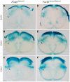
Red nucleus and rubrospinal tract disorganization in the absence of Pou4f1
- PMID: 25698939
- PMCID: PMC4318420
- DOI: 10.3389/fnana.2015.00008
Red nucleus and rubrospinal tract disorganization in the absence of Pou4f1
Abstract
The red nucleus (RN) is a neuronal population that plays an important role in forelimb motor control and locomotion. Histologically it is subdivided into two subpopulations, the parvocellular RN (pRN) located in the diencephalon and the magnocellular RN (mRN) in the mesencephalon. The RN integrates signals from motor cortex and cerebellum and projects to spinal cord interneurons and motor neurons through the rubrospinal tract (RST). Pou4f1 is a transcription factor highly expressed in this nucleus that has been related to its specification. Here we profoundly analyzed consequences of Pou4f1 loss-of-function in development, maturation and axonal projection of the RN. Surprisingly, RN neurons are specified and maintained in the mutant, no cell death was detected. Nevertheless, the nucleus appeared disorganized with a strong delay in radial migration and with a wider neuronal distribution; the neurons did not form a compacted population as they do in controls, Robo1 and Slit2 were miss-expressed. Cplx1 and Npas1, expressed in the RN, are transcription factors involved in neurotransmitter release, neuronal maturation and motor function processes among others. In our mutant mice, both transcription factors are lost, suggesting an abnormal maturation of the RN. The resulting altered nucleus occupied a wider territory. Finally, we examined RST development and found that the RN neurons were able to project to the spinal cord but their axons appeared defasciculated. These data suggest that Pou4f1 is necessary for the maturation of RN neurons but not for their specification and maintenance.
Keywords: Cplx1; Npas1; Pou4f1; development; maturation; midbrain; red nucleus; rubrospinal tract.
Figures


Similar articles
-
Postnatal maturation of the red nucleus motor map depends on rubrospinal connections with forelimb motor pools.J Neurosci. 2014 Mar 19;34(12):4432-41. doi: 10.1523/JNEUROSCI.5332-13.2014. J Neurosci. 2014. PMID: 24647962 Free PMC article.
-
Motor Cortex Activity Organizes the Developing Rubrospinal System.J Neurosci. 2015 Sep 30;35(39):13363-74. doi: 10.1523/JNEUROSCI.1719-15.2015. J Neurosci. 2015. PMID: 26424884 Free PMC article.
-
Somatotopic studies on red nucleus: spinal projection level and respective receptive fields.J Neurophysiol. 1975 Jul;38(4):965-80. doi: 10.1152/jn.1975.38.4.965. J Neurophysiol. 1975. PMID: 1159475
-
Red nucleus structure and function: from anatomy to clinical neurosciences.Brain Struct Funct. 2021 Jan;226(1):69-91. doi: 10.1007/s00429-020-02171-x. Epub 2020 Nov 12. Brain Struct Funct. 2021. PMID: 33180142 Free PMC article. Review.
-
Evolution of the red nucleus and rubrospinal tract.Behav Brain Res. 1988 Apr-May;28(1-2):9-20. doi: 10.1016/0166-4328(88)90072-1. Behav Brain Res. 1988. PMID: 3289562 Review.
Cited by
-
Developmental Requirement of Homeoprotein Otx2 for Specific Habenulo-Interpeduncular Subcircuits.J Neurosci. 2019 Feb 6;39(6):1005-1019. doi: 10.1523/JNEUROSCI.1818-18.2018. Epub 2018 Dec 28. J Neurosci. 2019. PMID: 30593496 Free PMC article.
-
Gli2-Mediated Shh Signaling Is Required for Thalamocortical Projection Guidance.Front Neuroanat. 2022 Feb 10;16:830758. doi: 10.3389/fnana.2022.830758. eCollection 2022. Front Neuroanat. 2022. PMID: 35221935 Free PMC article.
-
Celsr3 Inactivation in the Brainstem Impairs Rubrospinal Tract Development and Mouse Behaviors in Motor Coordination and Mechanic-Induced Response.Mol Neurobiol. 2022 Aug;59(8):5179-5192. doi: 10.1007/s12035-022-02910-7. Epub 2022 Jun 9. Mol Neurobiol. 2022. PMID: 35678978 Free PMC article.
-
Heterogeneous Diffusion Metrics Patterns in Delayed Encephalopathy After Acute Carbon Monoxide Poisoning: A Case Report.Brain Neurorehabil. 2023 Nov 6;16(3):e34. doi: 10.12786/bn.2023.16.e34. eCollection 2023 Nov. Brain Neurorehabil. 2023. PMID: 38047103 Free PMC article.
References
LinkOut - more resources
Full Text Sources
Other Literature Sources

