. 2015:2015:254237.
doi: 10.1155/2015/254237. Epub 2015 Jan 28.

Viability, apoptosis, proliferation, activation, and cytokine secretion of human keratoconus keratocytes after cross-linking

Affiliations

Viability, apoptosis, proliferation, activation, and cytokine secretion of human keratoconus keratocytes after cross-linking

Xuefei Song et al. Biomed Res Int. 2015.

Abstract

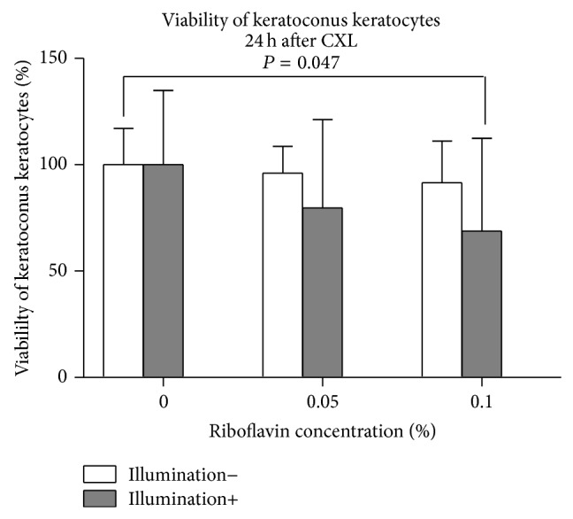
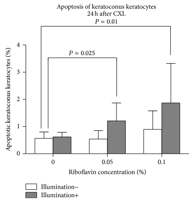
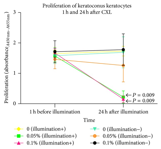
Purpose: The purpose of this study was to determine the impact of cross-linking (CXL) on viability, apoptosis, proliferation, activation, and cytokine secretion of human keratoconus (KC) keratocytes, in vitro.

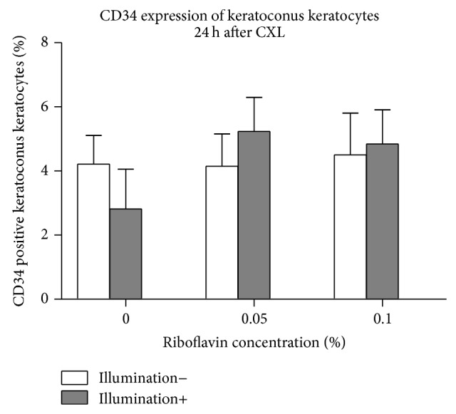
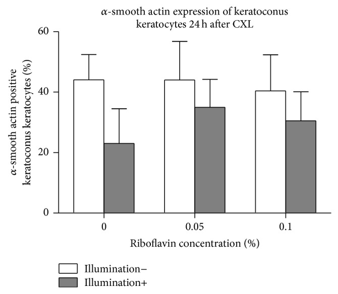
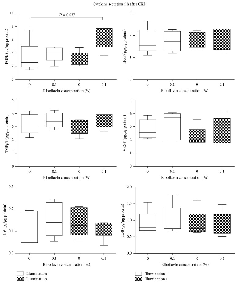
Methods: Primary KC keratocytes were cultured in DMEM/Ham's F12 medium supplemented with 10% FCS and underwent UVA illumination (370 nm, 2 J/cm(2)) during exposure to 0.1% riboflavin and 20% Dextran in PBS. Twenty-four hours after CXL, viability was assessed using Alamar blue assay; apoptosis using APO-DIRECT Kit; proliferation using ELISA-BrdU kit; and CD34 and alpha-smooth muscle actin (α-SMA) expression using flow cytometry. Five and 24 hours after CXL, FGFb, HGF, TGFβ1, VEGF, KGF, IL-1β, IL-6, and IL-8 secretion was measured using enzyme-linked-immunoabsorbent assay (ELISA).

Results: Following CXL, cell viability and proliferation decreased (P < 0.05; P = 0.009), the percentage of apoptotic keratocytes increased (P < 0.05) significantly, and CD34 and α-SMA expression remained unchanged (P > 0.06). Five hours after CXL, FGFb secretion increased significantly (P = 0.037); however no other cytokine secretion differed significantly from controls after 5 or 24 hours (P > 0.12).

Conclusions: Cross-linking decreases viability, triggers apoptosis, and inhibits proliferation, without an impact on multipotent hematopoietic stem cell transformation and myofibroblastic transformation of KC keratocytes. CXL triggers FGFb secretion of KC keratocytes transiently (5 hours), normalizing after 24 hours.


Figures

Figure 1
Figure 1
Viability of human KC keratocytes 24 hours following CXL. Application of 0.05% or 0.1% riboflavin-5-phosphate solution without illumination or the UVA-light illumination alone did not show a significant impact on keratocyte viability compared to controls (P > 0.34). Using 0.1% CXL, viability of KC keratocytes decreased significantly (P = 0.047).
Figure 2
Figure 2
Percentage of apoptotic KC keratocytes 24 hours following CXL. The use of riboflavin or UVA-light illumination separately had no significant impact on the percentage of apoptotic keratocytes compared to controls (P > 0.262). Following CXL using 0.05% or 0.1% riboflavin, the percentage of apoptotic KC keratocytes was significantly increased (P = 0.025 and P = 0.01, resp.).
Figure 3
Figure 3
Proliferation of human KC keratocytes 24 hours after CXL. Twenty-four hours after illumination, the proliferation of keratocytes was significantly inhibited using 0.05% or 0.1% riboflavin (P = 0.009 for both concentrations) compared to controls. Proliferation of keratocytes remained unchanged using riboflavin or illumination alone (P > 0.25). The 1 h before illumination group was tested as a starting point in cell growth under the same conditions.
Figure 4
Figure 4
CD34 expression of keratoconus keratocytes 24 hours after CXL. Using riboflavin or illumination only, expression of CD34 in KC keratocytes did not change significantly compared to controls (P > 0.873). Twenty-four hours after CXL, CD34 expression of keratocytes also remained unchanged at both riboflavin concentrations (P = 0.15 and P = 0.34), compared to controls.
Figure 5
Figure 5
α-SMA expression of KC keratocytes 24 hours after CXL. Twenty-four hours following treatment, there was no significant difference in the percentage of alpha-SMA positive KC keratocytes using riboflavin or UVA-light illumination separately (P > 0.16). Using CXL, α-SMA expression in keratocytes also remained unchanged at both riboflavin concentrations (P = 0.15 and P = 0.06), compared to controls.
Figure 6
Figure 6
Cytokine secretion 5 hours after cross-linking. Significant differences are indicated.
Figure 7
Figure 7
Cytokine secretion 24 hours after cross-linking. No significant difference could be detected compared to untreated controls.

References

    1. Romero-Jiménez M., Santodomingo-Rubido J., Wolffsohn J. S. Keratoconus: a review. Contact Lens & Anterior Eye. 2010;33(4):157–166. doi: 10.1016/j.clae.2010.04.006. - DOI - PubMed
    1. Rabinowitz Y. S. Keratoconus. Survey of Ophthalmology. 1998;42(4):297–319. doi: 10.1016/s0039-6257(97)00119-7. - DOI - PubMed
    1. Krachmer J. H., Feder R. S., Belin M. W. Keratoconus and related noninflammatory corneal thinning disorders. Survey of Ophthalmology. 1984;28(4):293–322. doi: 10.1016/0039-6257(84)90094-8. - DOI - PubMed
    1. Wollensak G., Spoerl E., Seiler T. Riboflavin/ultraviolet-A-induced collagen crosslinking for the treatment of keratoconus. American Journal of Ophthalmology. 2003;135(5):620–627. doi: 10.1016/s0002-9394(02)02220-1. - DOI - PubMed
    1. Raiskup F., Spoerl E. Corneal crosslinking with riboflavin and ultraviolet A. I. Principles. The Ocular Surface. 2013;11(2):65–74. doi: 10.1016/j.jtos.2013.01.002. - DOI - PubMed

Publication types

MeSH terms