. 2015 Feb 19;57(4):607-621.
doi: 10.1016/j.molcel.2015.01.021.

Pervasive and essential roles of the Top3-Rmi1 decatenase orchestrate recombination and facilitate chromosome segregation in meiosis

Affiliations

Pervasive and essential roles of the Top3-Rmi1 decatenase orchestrate recombination and facilitate chromosome segregation in meiosis

Shangming Tang et al. Mol Cell. .

Abstract

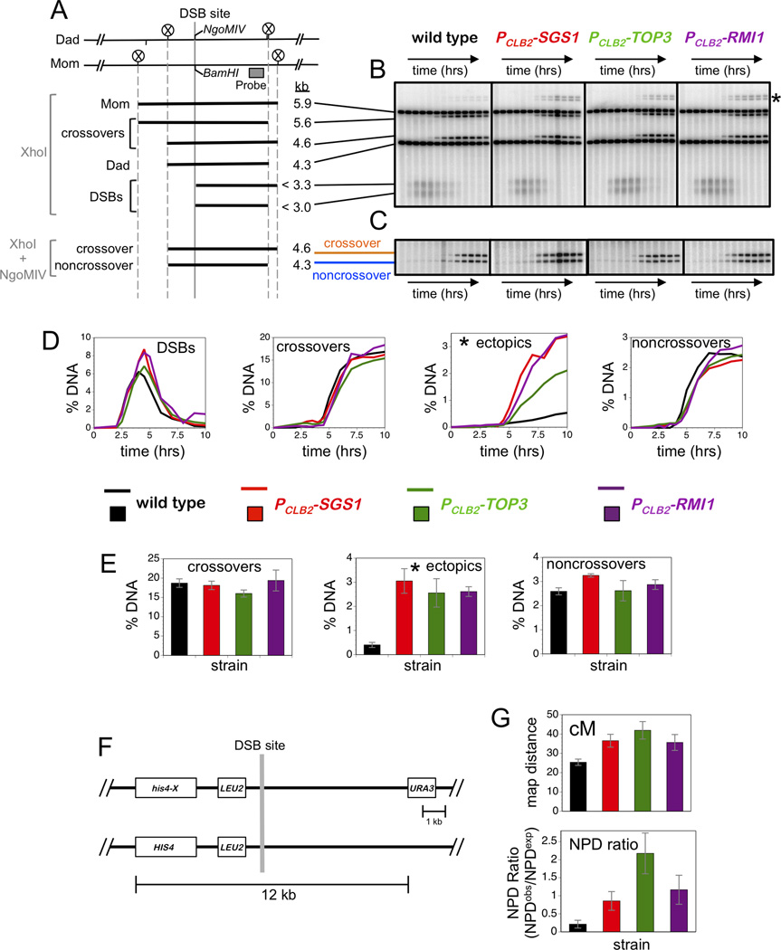
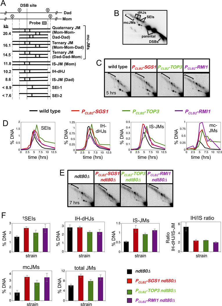
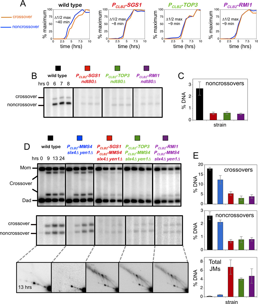
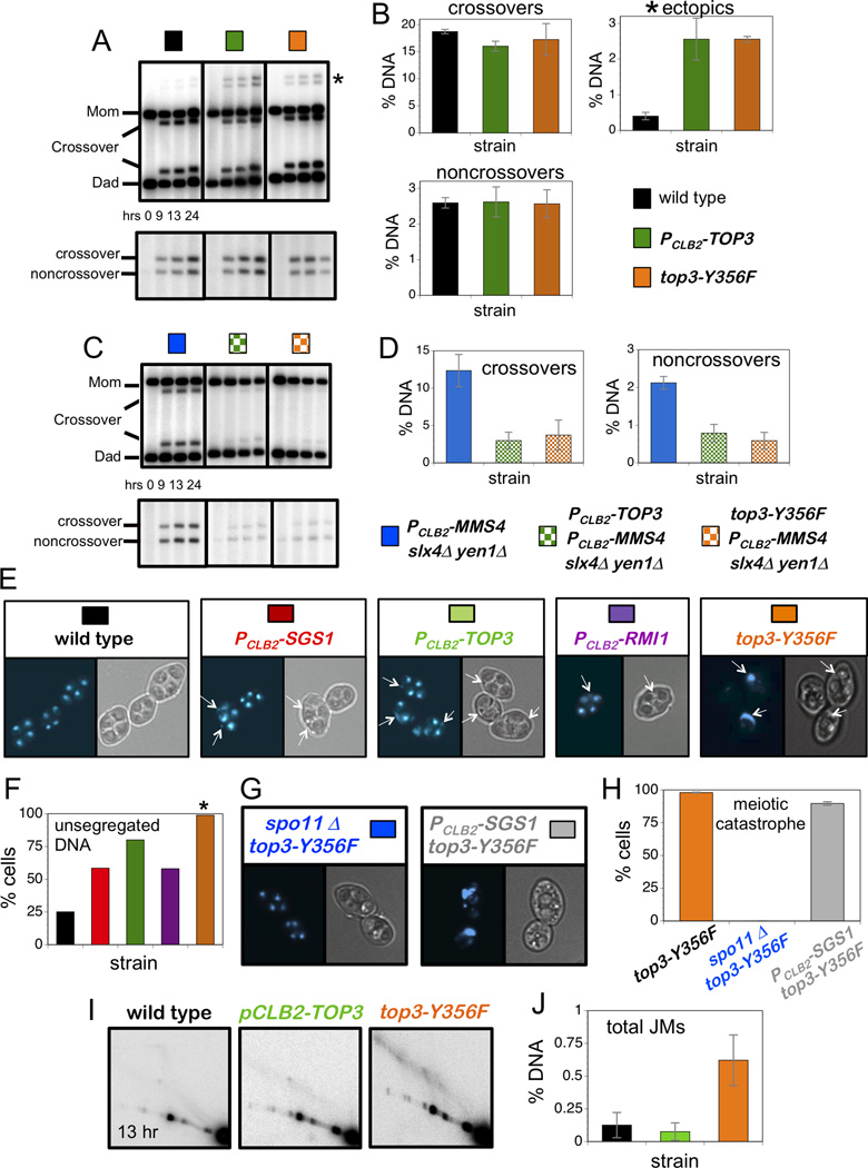
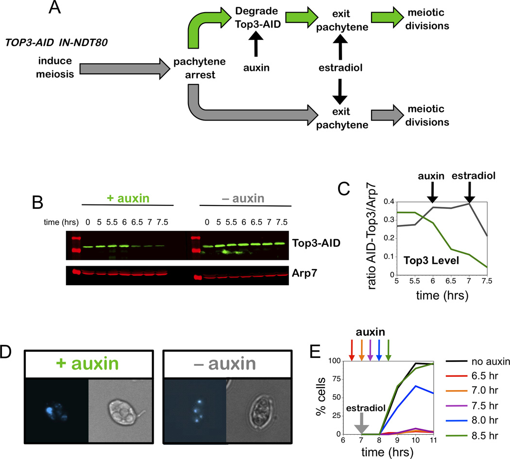
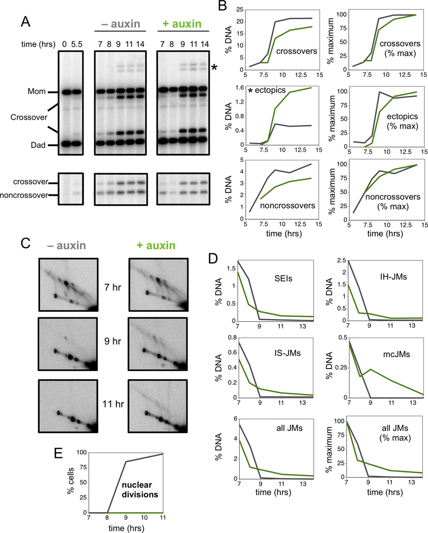
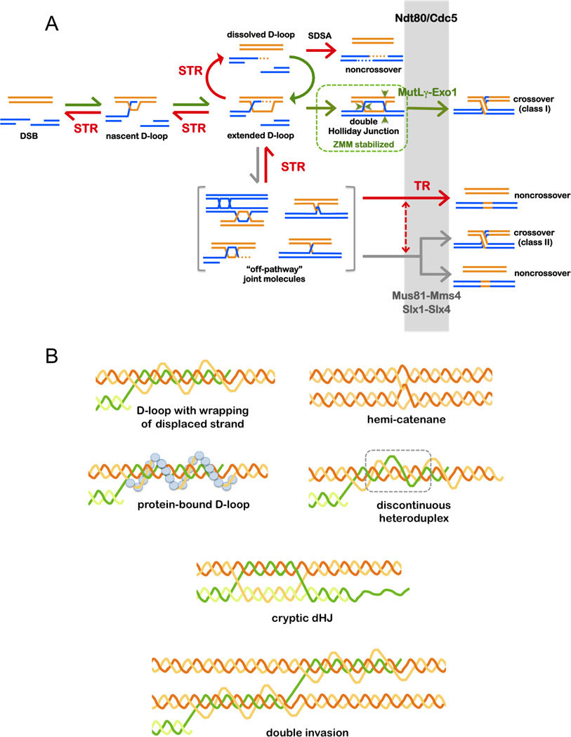
The Bloom's helicase ortholog, Sgs1, plays central roles to coordinate the formation and resolution of joint molecule intermediates (JMs) during meiotic recombination in budding yeast. Sgs1 can associate with type-I topoisomerase Top3 and its accessory factor Rmi1 to form a conserved complex best known for its unique ability to decatenate double-Holliday junctions. Contrary to expectations, we show that the strand-passage activity of Top3-Rmi1 is required for all known functions of Sgs1 in meiotic recombination, including channeling JMs into physiological crossover and noncrossover pathways, and suppression of non-allelic recombination. We infer that Sgs1 always functions in the context of the Sgs1-Top3-Rmi1 complex to regulate meiotic recombination. In addition, we reveal a distinct late role for Top3-Rmi1 in resolving recombination-dependent chromosome entanglements to allow segregation at anaphase. Surprisingly, Sgs1 does not share this essential role of Top3-Rmi1. These data reveal an essential and pervasive role for the Top3-Rmi1 decatenase during meiosis.


Figures

Figure 1
Figure 1. All STR Components are Required to Suppress Aberrant Recombination
(A) Map of the HIS4::LEU2 hotspot highlighting the DSB site, diagnostic restriction sites and position of the probe used in Southern analysis. Sizes of diagnostic fragments are shown below. Circled Xs indicate XhoI sites. (B) 1D Southern analysis of XhoI digested genomic DNA to monitor DSBs, allelic crossovers and ectopic crossovers (indicated by an asterisk). Time points are 0, 2, 2.5, 3, 3.5, 4, 4.5, 5, 6, 7, 8, 9, and 13 hours. (C) 1D Southern analysis of XhoI+NgoMIV doubly digested genomic DNA to monitor noncrossover formation. Samples are from the same time courses shown in panel B. (D) Quantitation of images shown in panels B and C. % DNA is percentage of total hybridizing DNA signal. (E) Quantification of allelic crossovers, ectopic crossovers and noncrossovers after 13 hrs from multiple independent time-courses. (F) Map of HIS4::LEU2 hotspot showing flanking genetic markers. (G) Map distances (upper panel) and NPD ratios (lower panel) for the interval shown in F. Data with error bars represent mean ± SEM. See also Figure S1 and Table S1.
Figure 2
Figure 2. Altered Joint Molecule Spectra in PCLB2-TOP3 and PCLB2-RMI1 Mutants
(A) JM structures detected at HIS4::LEU2, from lowest to highest mobility. Positions of diagnostic XhoI sites (circled Xs) and the Southern probe are shown. IS-JM, inter-sister joint molecule; IH-dHJ, inter-homolog double Holiday junction; SEI, single end invasion; mc-JMs, 3-and 4-chromatid joint molecules. (B) Southern blot of native/native 2D gel showing JMs detailed in panel A. (C) 2D Southern analysis of JMs in wild type, PCLB2-SGS1, PCLB2-TOP3 and PCLB2-RMI1 strains. (D) Quantification of individual JM species. Percent DNA is percent of total hybridization signal. (E) 2D Southern analysis of JMs in ndt80Δ, PCLB2-SGS1 ndt80Δ, PCLB2-TOP3 ndt80Δ and PCLB2-RMI1 ndt80Δ strains. (F) Quantification of individual JM species in ndt80Δ strains, total JMs and the ratio of IH-dHJs/IS-JMs. Percent DNA is percent of total hybridization signal. Data with error bars represent mean ± SEM.
Figure 3
Figure 3. Crossovers and Noncrossovers in the PCLB2-TOP3 and PCLB2-RMI1 Mutants Require the Structure-Selective Endonucleases
(A) Normalized curves to compare the timing of crossovers and noncrossovers from the time-courses analysis shown in Figure 1C. Δ1/2 max is the difference between the times of the half maximum values. (B) Representative Southern images of noncrossover analysis in the indicated ndt80Δ strains. (C) Quantification of noncrossovers in ndt80Δ strains at 7 hrs. (D) Representative Southern images of crossover (upper panel), noncrossover (middle panel) and JM analysis (bottom panel) at indicated times. (E) Quantification of crossovers, noncrossovers and total JMs at 13 hrs. Data with error bars represent mean ± SEM. See also Figures S2, S3 and S4.
Figure 4
Figure 4. Single-Strand Decatenase Activity of Top3 Suppresses Aberrant Recombination and is Essential for Chromosome Segregation
(A) Representative Southern images of crossover (upper panel) and noncrossover (lower panel) analysis in indicated strains. Asterisk indicates ectopic crossover bands. (B) Quantification of crossovers, ectopic recombination and noncrossovers at 13hrs. (C) Representative Southern images of crossover (upper panel) and noncrossover (lower panel) analysis in indicated strains. (D) Quantification of crossovers and noncrossovers at 13hrs. (E) Representative images of cells from indicated strains at 13 hrs. DAPI-stained and brightfield images of the same cells are shown. Arrows indicate unsegregated DNA masses. (F) Quantification of cells containing unsegregated DNA masses. Colors correspond to the strains shown in panel E. (G) Representative cells from spo11Δ top3-Y356F and PCLB2-SGS1 top3-Y356F strain at 13 hrs. (H) Quantification of cells showing meiotic catastrophe in top3-Y356F, spo11Δ top3-Y356F and PCLB2-SGS1 top3-Y356F strains. (I) Representative Southern images of 2D JM analysis at 13hrs. (J) Quantification of total JM levels at 13 hrs in the strains shown in panel I. Data with error bars represent mean ± SEM. See also Figure S4.
Figure 5
Figure 5. Top3 Acts Late in Meiotic Prophase to Promote Chromosome Separation
(A) Regimen to inactivate Top3-AID during late stages of meiotic prophase. (B) Western images showing Top3-AID levels in cells with and without addition of auxin at 6 hrs. Arp7 was used as loading control. (C) Quantification of Top3 levels from the experiment shown in panel B. (D) Representative images of cells from subcultures with and without addition of auxin at 6 hrs and addition of estradiol at 7 hrs, sampled at 11 hrs. DAPI-stained and brightfield images of the same cells are shown. (E) Quantification of nuclear divisions in subcultures with and without auxin treatment at indicated times, following induction of Ndt80 expression at 7 hrs. See also Figure S6.
Figure 6
Figure 6. Late Action of Top3 is Required for Efficient Recombination and Suppression of Ectopic Crossovers
(A) Southern images of crossover (upper panels) and noncrossover (lower panels) analysis from cell subcultures with and without auxin treatment (Top3-AID degradation) at 6.5 hrs and induction of Ndt80 expression at 7 hrs. (B) Quantification of allelic and ectopic crossovers, and noncrossovers from the experiment shown in panel A. Left panels show recombinants as percentage of total hybridizing DNA signal. Right panels show normalized curves to compare the timing of recombinants. (C) Southern blot images of 2D JM analysis. (D) Quantification of individual JM species, total JM levels and normalized JM levels in cells with and without auxin treatment. (E) Quantification of nuclear divisions in the two subcultures. See also Figure S7.
Figure 7
Figure 7. Roles of the Top3-Rmi1 Single-Strand Decatenase in Meiosis
(A) Pathways of meiotic recombination highlighting the prevalent role of STR’s compound helicase-decatenase activity in regulating JM formation; and the late role of the TR decatenase in removing recombination-dependent entanglements. STR dissolves nascent and extended D-loops to reset DSBs. This process will reverse unproductive and excessive strand exchanges allowing DSBs to either engage in new rounds of strand invasion, or to anneal. Annealing can occur either as part of the SDSA process to form Ndt80/Cdc5-independent noncrossovers, or during second-end capture to form dHJ crossover precursors (Lao et al., 2008). The latter involves stabilization by ZMM proteins, such as MutSγ (Msh4-Msh5), which protect crossover-designated dHJs from STR-mediated dissolution. Crossover-specific resolution requires Cdc5/Plk1 and the Exo1-MutLγ ensemble. By dissolving nascent JMs, STR limits accumulation of off-pathway JMs that include mcJMs, trapped and dead-end intermediates, topologically-entrapped JMs, and structures that require resolution by the structure-selective endonucleases. Resolution of such structures involves Cdc5/Plk1, which generally activates JM resolution and directly activates Mus81-Mms4 (Matos et al., 2011). By promoting disassembly of synaptonemal complexes, Cdc5/Plk1 may also expose trapped JMs to the TR decatenase and the structure-selective endonucleases. A subset of noncrossovers derive from Cdc5/Plk1-dependent dissolution by TR. Resolution of some off-pathway JMs may require collaboration of TR and the structure-selective endonucleases, as indicated by the dashed arrow. At late stages, the TR decatenase may act broadly to remove residual entanglements from all JMs. (B) Potential topological constraints and complexities in meiotic JMs that might necessitate a general requirement for the Top3-Rmi1 single-strand decatenase.

Comment in

  • TOPping off meiosis.
    Haber JE. Haber JE. Mol Cell. 2015 Feb 19;57(4):577-581. doi: 10.1016/j.molcel.2015.02.004. Mol Cell. 2015. PMID: 25699706 Free PMC article.

References

    1. Allers T, Lichten M. Differential timing and control of noncrossover and crossover recombination during meiosis. Cell. 2001;106:47–57. - PubMed
    1. Andersen SL, Sekelsky J. Meiotic versus mitotic recombination: two different routes for double-strand break repair: the different functions of meiotic versus mitotic DSB repair are reflected in different pathway usage and different outcomes. BioEssays : news and reviews in molecular, cellular and developmental biology. 2010;32:1058–1066. - PMC - PubMed
    1. Bachrati CZ, Borts RH, Hickson ID. Mobile D-loops are a preferred substrate for the Bloom's syndrome helicase. Nucleic acids research. 2006;34:2269–2279. - PMC - PubMed
    1. Bell L, Byers B. Separation of branched from linear DNA by two-dimensional gel electrophoresis. Anal Biochem. 1983;130:527–535. - PubMed
    1. Bellendir SP, Sekelsky J. An elegans Solution for Crossover Formation. PLoS genetics. 2013;9:e1003658. - PMC - PubMed

Publication types

MeSH terms

Substances