. 2015 Feb 19;57(4):636-647.
doi: 10.1016/j.molcel.2015.01.011.

BRCA1 recruitment to transcriptional pause sites is required for R-loop-driven DNA damage repair

Affiliations

BRCA1 recruitment to transcriptional pause sites is required for R-loop-driven DNA damage repair

Elodie Hatchi et al. Mol Cell. .

Abstract

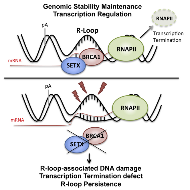
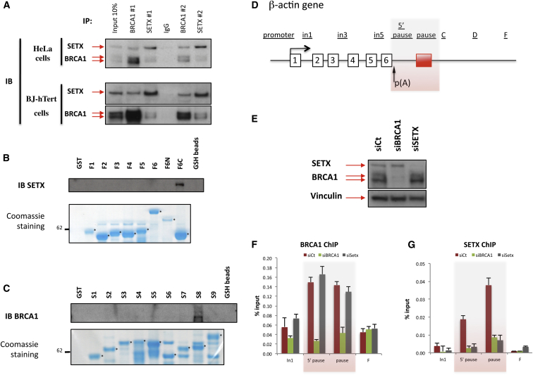
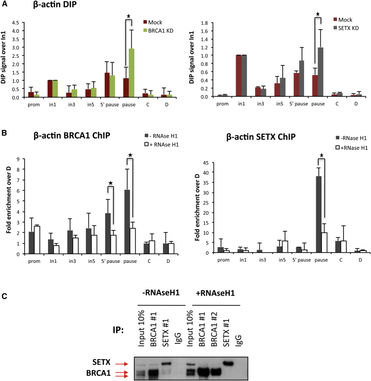
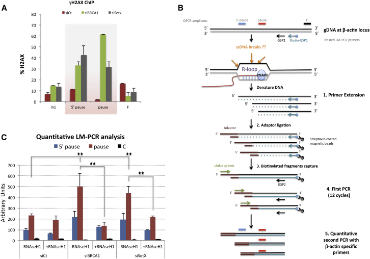
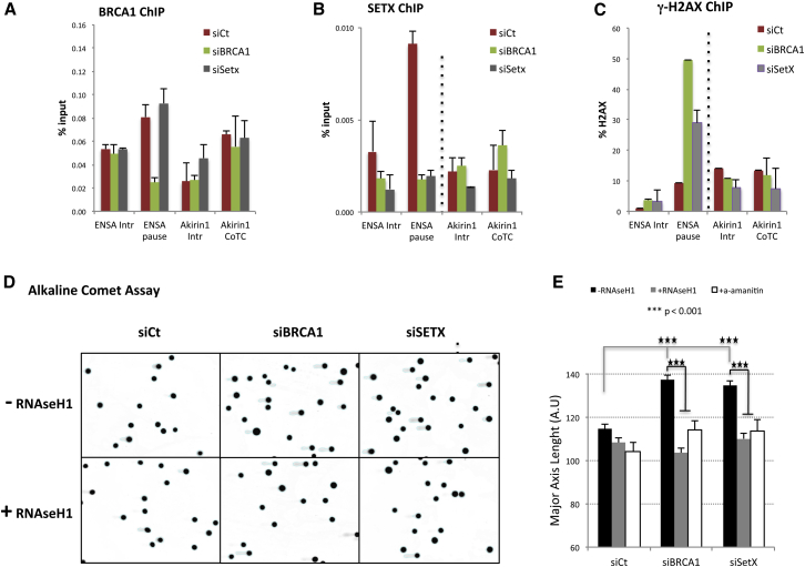
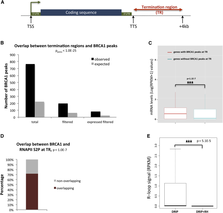
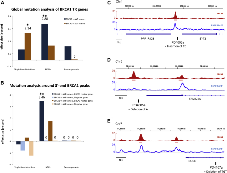
The mechanisms contributing to transcription-associated genomic instability are both complex and incompletely understood. Although R-loops are normal transcriptional intermediates, they are also associated with genomic instability. Here, we show that BRCA1 is recruited to R-loops that form normally over a subset of transcription termination regions. There it mediates the recruitment of a specific, physiological binding partner, senataxin (SETX). Disruption of this complex led to R-loop-driven DNA damage at those loci as reflected by adjacent γ-H2AX accumulation and ssDNA breaks within the untranscribed strand of relevant R-loop structures. Genome-wide analysis revealed widespread BRCA1 binding enrichment at R-loop-rich termination regions (TRs) of actively transcribed genes. Strikingly, within some of these genes in BRCA1 null breast tumors, there are specific insertion/deletion mutations located close to R-loop-mediated BRCA1 binding sites within TRs. Thus, BRCA1/SETX complexes support a DNA repair mechanism that addresses R-loop-based DNA damage at transcriptional pause sites.


Figures

None
Graphical abstract
Figure 1
Figure 1
BRCA1 Interacts with SETX and Is Required for SETX Recruitment to the R-Loop-Associated Termination Pause Region of the Human β-actin Gene (A) Co-immunoprecipitation (IP) of endogenous BRCA1 and SETX in HeLa cell (top) and in BJ-hTert human fibroblast extracts (bottom), using antibodies against BRCA1 (BRCA1 #1/#2) or SETX (SETX #1/#2). IgG, negative control. (B) In vitro interaction assay using recombinant GST-BRCA1 fragments performed in HeLa cells. GSH bead-bound proteins were eluted and analyzed by immunoblot. Bottom: relative abundance of each recombinant affinity purified fusion protein (marked with a star) visualized by SDS-PAGE after Coomassie staining. Additional bands are readthrough and/or degradation products of recombinant proteins. (C) Same as in (B) but with recombinant GST-SETX fragments. (D) Schematic representation of the human β-actin gene: exons are numbered, and the red box reflects the existence of a transcription pause element. Location of primers used for the ChIP experiments are depicted above the diagram. (E) Immunoblot that reflects the efficiency of the siRNA-mediated depletion of BRCA1 and SETX. Top: SETX and BRCA1; bottom: Vinculin used as a loading control. (F) ChIP analyses performed on the β-actin gene from mock-treated (red bars), BRCA1- (green bars), or SETX-depleted cells (gray bars) using BRCA1 Ab #3. Average ChIP values ± SD from three independent experiments are shown. (G) Same ChIP experiments as in (F) but with SETX Ab #1. See also Figures S1 and S2.
Figure 2
Figure 2
BRCA1 Recruitment to the β-actin Termination Pause Site Is Mediated by R-Loops (A) RNA:DNA immunoprecipitation (DIP) analyses performed in HeLa from mock-treated or BRCA1-depleted cells (BRCA1 KD) (left) and mock-treated or SETX-depleted cells (SETX KD) (right). (B) BRCA1 and SETX ChIP experiment performed in control conditions (−RNaseH1) or with RNaseH1 overexpression (+RNaseH1). Average DIP and ChIP values ± SD from three, independent experiments are shown. p < 0.05. (C) BRCA1 and SETX co-IP experiments performed in HeLa cells ± RNaseH1 expression. IgG, negative control.
Figure 3
Figure 3
BRCA1/SETX Complex at the β-actin Pause Site Protects Cells from R-Loop-Driven ssDNA Breaks (A) ChIP analysis performed on the β-actin gene as in Figure 1 using γH2AX and total H2AX antibodies. Histograms represent the proportion of total H2AX phosphorylated on Ser139 (i.e., γH2AX). Average ChIP values ± SD from three independent experiments are shown. (B) LM-PCR strategy used to identify R-loop-associated ssDNA breaks on the coding strand. See Supplemental Experimental Procedures. (C) Quantitative detection of ssDNA after LM-PCR performed on the β-actin gene before and after BRCA1 or SETX knockdown and with or without ectopic RNaseH1 expression. QPCR values are average ± SD from three independent experiments. p < 0.05, ∗∗p < 0.007 by one-tailed Student’s t test. See also Figure S3.
Figure 4
Figure 4
DNA Damage Arising in Absence of BRCA1/SETX Complexes at Termination Pause Sites Is R-Loop Dependent (A and B) BRCA1 (A) and SETX (B) ChIP analyses performed on the ENSA and Akirin1 genes in HeLa cells transfected with siCt, siBRCA1, or siSETX. ENSA and Akirin1 transcription termination is regulated by R-loops and CoTC sequences, respectively. Intronic regions (Intr) were studied as controls. Average ChIP values ± SD from three independent experiments are shown. (C) γH2AX ChIP experiments performed as in (A) and analyzed as in Figure 3A. Average ChIP values ± SD from three independent experiments are shown. (D) Representative pictures of comet assays performed under alkaline conditions in HeLa cells transfected with siCt, siBRCA1, or siSETX in the absence (−RNaseH1) and presence (+RNaseH1) of ectopic RNaseH1 expression. (E) Quantitative analysis of comet tail lengths for each condition showed in (D). Average tail lengths ±SEM from three independent experiments are shown. ∗∗∗p < 0.001 by two-tailed Student’s t test. See also Figure S4.
Figure 5
Figure 5
BRCA1 Binds the Transcription Termination Region of a Substantial Subset of Actively Expressed Mammalian Genes (A) Diagram of candidate BRCA1 TR binding regions. Putative termination regions (TRs) were defined as segments extending from the TTS to TTS + 4 kb. (B) Total number of observed and expected overlaps between TR and BRCA1 peaks (ppoiss = 2.2E-177). “Filtered”: BRCA1 ChIP-seq peaks divested of those overlapping promoter regions and transcripts (ppoiss = 1.2E-42). “Expressed filtered”: BRCA1 peaks present in TR region of expressed genes (ppoiss = 5E-24). (C) Gene expression comparison between genes with (red) and without (blue) BRCA1 bound to relevant TR. Boxplots reflect the median (50th percentile) of mRNA expression. ∗∗∗p = 1.1E-7, Mann-Whitney test. Outliers have been omitted from the plot. (D) Overlap between BRCA1 TR and RNAPIISer2P peaks, overlap enrichment over random (∗∗∗p < 1.0E-7), see Experimental Procedures. (E) Boxplots showing DRIP-seq signals (RPKM) of DRIP samples compared with DRIP+RH controls (treated with RNaseH1) in BRCA1 TR candidate regions, ∗∗∗p = 5.101E-5, paired Wilcoxon test. See also Figure S5 and Table S1.
Figure 6
Figure 6
BRCA1-Deficient Breast Cancers Reveal Genomic Abnormalities at and near BRCA1-Associated Termination Sites (A and B) Global mutational analysis carried out in the 184 BRCA1 TR genes using the complete whole-genome catalog of somatic mutations from 21 breast cancers (Nik-Zainal et al., 2012). Effect size comparison (one-tailed CMH Z score) between the different tumor subgroups when testing the region from TSS − 1,250 bp to TTS + 5 kb (A) or ± 4 kb from TTS (B). WT tumors = non-BRCA1/BRCA2, and negative genes = CoTC genes. Statistical significant: p < 0.05 and ∗∗p < 0.01. (C–E) ChIP-seq profiles of BRCA1 (red) and RNAPIISer2P (blue) in BRCA1 TR genes and location of indels (black boxes). Chr, chromosome. See also Figure S6 and Table S2.

Similar articles

Cited by

References

    1. Aguilera A. The connection between transcription and genomic instability. EMBO J. 2002;21:195–201. - PMC - PubMed
    1. Aguilera A., García-Muse T. R loops: from transcription byproducts to threats to genome stability. Mol. Cell. 2012;46:115–124. - PubMed
    1. Alexandrov L.B., Nik-Zainal S., Wedge D.C., Aparicio S.A.J.R., Behjati S., Biankin A.V., Bignell G.R., Bolli N., Borg A., Børresen-Dale A.-L., Australian Pancreatic Cancer Genome Initiative. ICGC Breast Cancer Consortium. ICGC MMML-Seq Consortium. ICGC PedBrain Signatures of mutational processes in human cancer. Nature. 2013;500:415–421. - PMC - PubMed
    1. Alzu A., Bermejo R., Begnis M., Lucca C., Piccini D., Carotenuto W., Saponaro M., Brambati A., Cocito A., Foiani M., Liberi G. Senataxin associates with replication forks to protect fork integrity across RNA-polymerase-II-transcribed genes. Cell. 2012;151:835–846. - PMC - PubMed
    1. Anderson S.F., Schlegel B.P., Nakajima T., Wolpin E.S., Parvin J.D. BRCA1 protein is linked to the RNA polymerase II holoenzyme complex via RNA helicase A. Nat. Genet. 1998;19:254–256. - PubMed

Publication types