Review
. 2014:79:81-92.
doi: 10.1101/sqb.2014.79.024786. Epub 2015 Feb 19.

Internal and External Feedback Circuits for Skilled Forelimb Movement

Affiliations
Review

Internal and External Feedback Circuits for Skilled Forelimb Movement

Eiman Azim et al. Cold Spring Harb Symp Quant Biol. 2014.

Abstract

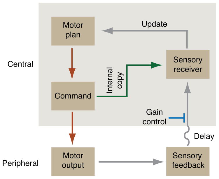
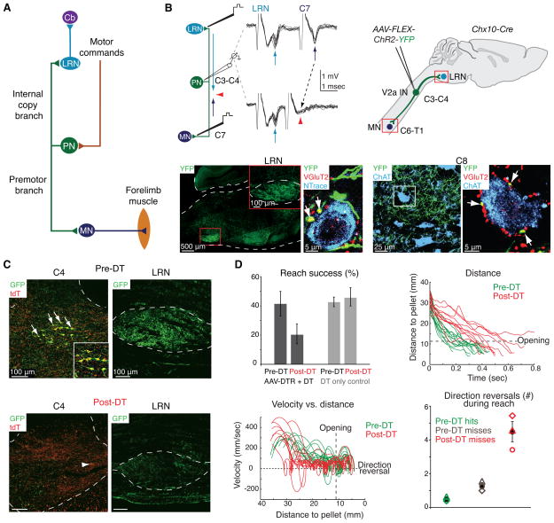
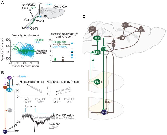
Skilled motor behavior emerges from interactions between efferent neural pathways that induce muscle contraction and feedback systems that report and refine movement. Two broad classes of feedback projections modify motor output, one from the periphery and a second that originates within the central nervous system. The mechanisms through which these pathways influence movement remain poorly understood, however. Here we discuss recent studies that delineate spinal circuitry that binds external and internal feedback pathways to forelimb motor behavior. A spinal presynaptic inhibitory circuit regulates the strength of external feedback, promoting limb stability during goal-directed reaching. A distinct excitatory propriospinal circuit conveys copies of motor commands to the cerebellum, establishing an internal feedback loop that rapidly modulates forelimb motor output. The behavioral consequences of manipulating these two circuits reveal distinct controls on motor performance and provide an initial insight into feedback strategies that underlie skilled forelimb movement.


Figures

Figure 1
Figure 1
Internal and external feedback motor pathways. During limb movement, motor plans are translated into commands by supraspinal and spinal pathways, eliciting motor output in the form of muscle contraction. Movement generates sensory feedback, including proprioceptive feedback from muscles, which conveys information about motor outcome to central sensory recipient centers. Temporal delays in sensory feedback imply a need for: a constraint on sensory feedback gain (blue) to maintain stability; and a more rapid mechanism of internal copy feedback (green) for online updating and correction of movement. Adapted from (Azim 2014).
Figure 2
Figure 2
Reaching kinematics in mice. (A) Mice were trained to reach for a food pellet through an opening in a transparent box. High-speed, high-resolution video capture and automated tracking of an infrared-reflective marker attached to the right paw were used to reconstruct three-dimensional reaching kinematics. (B) Successful (defined as retrieval of the food pellet) and unsuccessful reaches are highly stereotyped in wildtype mice. Trajectories of successful (hits, green traces) and unsuccessful (misses, brown traces) trials from a representative mouse are shown on the left. Paw velocity as a function of distance to the pellet is shown on the right. The transition from the reach phase to the late grab phase of movement is delineated by the box opening (vertical dashes). Velocity crossings of zero (horizontal dashes) indicate paw direction reversals. Adapted from (Azim et al. 2014).
Figure 3
Figure 3
Gad2-expressing neurons mediate presynaptic inhibition. (A) Proprioceptive sensory neurons (SN) convey sensory feedback signals from muscle to motor neurons (MN). Presynaptic inhibitory (GABApre) neurons contact sensory afferent terminals, while postsynaptic inhibitory (GABApost) neurons contact motor neurons directly. GABApre neurons express Gad2 (top schematic and left two images; far left: red Gad2ON contacts on blue VGluT1+ sensory afferent terminal, adjacent green GFP+ motor neuron labeled in Hb9-GFP mice). GABApost neurons express Gad1 (bottom schematic and right two images; second from right: red Gad1ON contacts adjacent green GFP+ motor neuron). While GABApre neurons express both Gad1 and Gad2 (second image from left: yellow Gad1ON/Gad2ON puncta adjacent VGluT1+ sensory terminal), GABApost neurons express Gad1 alone (far right image: red Gad1ON/Gad2OFF puncta). (B) Injection of Cre-dependent virus (AAV-FLEX-ChR2-YFP) into cervical cord of adult Gad2-Cre mice labels GABApre neurons (top, YFP+, Gad2+ contact adjacent VGluT1+ sensory terminal) and not GABApost neurons (bottom, YFP-negative red puncta). Viral injection marks ~80% of GABApre and ~1% of GABApost boutons. (C) After AAV-FLEX-ChR2-YFP injection in lumbar spinal cord of neonatal Gad2-Cre mice, recordings in isolated spinal cord from sensory afferents (dorsal root L4, extracellular) reveal primary afferent depolarization and, (D) recordings from motor neurons (whole cell patch clamp) reveal suppression of monosynaptic sensory-evoked excitatory postsynaptic currents after photostimulation (black, control; blue, 473 nm wavelength (λ) photostimulation). At early ages Gad2 is also expressed in GABApost boutons; therefore, all behavioral experiments were performed after viral injection in adult mice, when Gad2 expression is specific for GABApre neurons (B) (see (Fink et al. 2014) for details). Adapted from (Betley et al. 2009; Fink et al. 2014).
Figure 4
Figure 4
GABApre neuron ablation results in forelimb oscillation during reaching. (A) Cervical GABApre interneurons (INs) were targeted for acute ablation via injection of a conditional virus expressing a diphtheria toxin receptor-GFP fusion (AAV-FLEX-DTR-GFP) in cervical cord of adult Gad2-Cre mice. Before diphtheria toxin (DT) administration (left), ~90% of VGluT1+ sensory afferent terminals (blue) are contacted by Gad2+ GABApre boutons (red). Following DT administration (right), Gad2+ GABApre boutons contact <10% of sensory afferent terminals. Error bars indicate s.e.m. (B) GABApre ablation severely affects reach success; before (green) and after (red) DT administration for three Gad2-Cre mice. (C) Paw velocity as a function of time for a representative mouse (thin lines, individual reaches; bold lines, mean) before (green) and after (red) DT administration reveals severe oscillations after GABApre ablation. (D) Average power spectra (left, bold lines, mean across mice; shading, s.e.m.) of pre-DT (green) and post-DT (red) reaches indicate a narrow peak in oscillation frequency focused at ~20 Hz after ablation. Alignment of individual reaches to the point of maximal reach velocity (right) reveals consistent oscillatory decay in individual mice (faint red lines) and averaged across mice (bold red line). Fitting single exponential functions to the first three oscillatory peaks (gray dots and faint gray lines, individual mice; black line, mean) reveals a decay time of ~77 msec across mice. (E) A simplified model of a joint with sensory feedback controlled by a flexor (Tf) and extensor (Te) torque and defined by joint angle θ (left panels). Sensory feedback (s) depends on joint angle and normally permits smooth joint extension (gray arrows, black control trace) in the presence of external extensor drive (Ie). In the absence of presynaptic inhibition (PSI), simulated by imposing high sensory gain (h), sensory feedback triggers oscillation (red arrows and red high gain trace). Oscillation frequency and decay time change little across a wide range of gain values in the model (right plots), providing a possible explanation for why frequency and decay time are so consistent across mice (D). Gray shading indicates the range of experimentally observed peak oscillation frequency and decay times in post-DT Gad2-Cre mice, as well as the corresponding range of normalized gain values. Adapted from (Fink et al. 2014).
Figure 5
Figure 5
V2a PN ablation disrupts reaching movements. (A) PNs receive input from descending motor command pathways. PN axons bifurcate to innervate forelimb motor neurons (MN, premotor branch) and the lateral reticular nucleus (LRN, internal copy branch), which projects to the cerebellum (Cb). (B) In vivo extracellular recording in mouse cervical spinal cord while electrically stimulating LRN and C7 ventral horn reveals neurons fired antidromically from both stimulation sites (top left, arrows). Spike collision (red arrowheads) shows that the identified neuron projects to both targets, confirming the existence of PNs in mice. Injection of Cre-dependent virus (AAV-FLEX-ChR2-YFP) into cervical cord of adult Chx10-Cre mice labels V2a interneurons (IN) selectively (top right). YFP+ V2a INs project to the LRN (bottom left), where they form VGluT2+ excitatory synaptic contacts (red, arrows) onto LRN neurons (blue). Similarly, forelimb motor neurons in C8 (bottom right, blue) receive VGluT2+ (red), YFP+ contacts (arrows). (C) Cervical V2a INs were targeted for acute ablation via injection of a conditional virus expressing a diphtheria toxin receptor-GFP fusion (AAV-FLEX-DTR-GFP) in cervical cord of adult Chx10-Cre mice (top left, arrows, V2a INs genetically labeled by tdT in red; top right, GFP+ projections to the LRN). Diphtheria toxin (DT) administration ablates ~85% of V2a INs (bottom left, arrowhead indicates spared neuron), and eliminates GFP+ PN projections to the LRN (bottom right). (D) V2a IN elimination reduces reaching success (top left), and increases movement duration (top right) and the number of forelimb direction reversals (bottom left and right) during the reaching phase of movement (before the box opening). Shapes in bottom right plot represent individual mice. Error bars indicate s.e.m. Locomotion and digit abduction were not affected by V2a IN elimination (not shown). Adapted from (Azim et al. 2014).
Figure 6
Figure 6
PN internal copy activation modulates forelimb movement through a rapid cerebellar-motor loop. (A) ChR2 was expressed in cervical V2a INs via injection of conditional virus (AAV-FLEX-ChR2-YFP) in cervical cord of adult Chx10-Cre mice. Photostimulation of ChR2+ PN terminals in the LRN activates the PN-LRN pathway selectively, without eliciting antidromic action potentials (not shown). Photostimulation disrupts reaching movements (blue traces), producing a large increase in the incidence of paw direction reversals and variability in velocity and acceleration during reaching, without affecting digit abduction (not shown). Shapes in right plot represent individual mice. Error bars indicate s.e.m. (B) Motor field potential recordings in cervical spinal segments, as well as intracellular motor neuron recordings (not shown), reveal rapid activation of forelimb motor neurons (earliest activation ~4–5 msec from light onset). Severing the inferior cerebellar peduncles (ICP), which contain the projections from the LRN to the cerebellum, reduces the amplitude of motor field potentials by up to 65% and eliminates the fastest motor field onsets, implicating a rapid cerebellar-motor feedback pathway. (C) A putative subcortical PN-LRN feedback loop (green) recruits deep cerebellar nuclei (DCN), which excite reticulospinal (RS) projections to motor neurons (MN). Longer loops (brown) might engage cerebellar cortex (Cb Ctx), rubrospinal neurons (RN) and cortical pathways in posterior parietal cortex (PPC) and motor cortex (MC) via thalamus (Th) (see text). (A,B) adapted from (Azim et al. 2014).

References

    1. Ali MS, Hou ZK, Noori MN. Stability and performance of feedback control systems with time delays. Computers & Structures. 1998;66:241–248.
    1. Alstermark B, Ekerot CF. The Lateral Reticular Nucleus: a precerebellar center providing the Cerebellum with overview and integration of motor functions at systems level. A new hypothesis. The Journal of physiology. 2013;591:5453–5458. - PMC - PubMed
    1. Alstermark B, Isa T. Circuits for skilled reaching and grasping. Annual review of neuroscience. 2012;35:559–578. - PubMed
    1. Alstermark B, Kummel H, Pinter MJ, Tantisira B. Integration in descending motor pathways controlling the forelimb in the cat. 17. Axonal projection and termination of C3-C4 propriospinal neurones in the C6-Th1 segments. Experimental brain research Experimentelle Hirnforschung Experimentation cerebrale. 1990;81:447–461. - PubMed
    1. Alstermark B, Lindstrom S, Lundberg A, Sybirska E. Integration in descending motor pathways controlling the forelimb in the cat. 8. Ascending projection to the lateral reticular nucleus from C3-C4 propriospinal also projecting to forelimb motoneurones. Experimental brain research Experimentelle Hirnforschung Experimentation cerebrale. 1981a;42:282–298. - PubMed

Publication types

LinkOut - more resources