. 2015 Feb 20;347(6224):882-6.
doi: 10.1126/science.aaa1823.

Kinase dynamics. Using ancient protein kinases to unravel a modern cancer drug's mechanism

Affiliations

Kinase dynamics. Using ancient protein kinases to unravel a modern cancer drug's mechanism

C Wilson et al. Science. .

Abstract

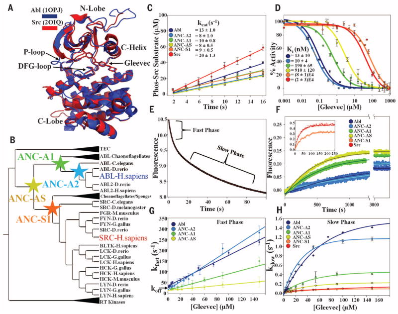
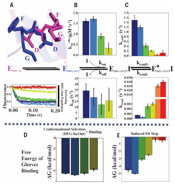
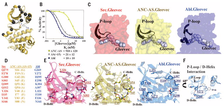
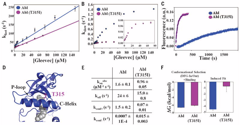
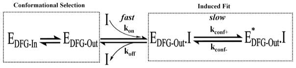
Macromolecular function is rooted in energy landscapes, where sequence determines not a single structure but an ensemble of conformations. Hence, evolution modifies a protein's function by altering its energy landscape. Here, we recreate the evolutionary pathway between two modern human oncogenes, Src and Abl, by reconstructing their common ancestors. Our evolutionary reconstruction combined with x-ray structures of the common ancestor and pre-steady-state kinetics reveals a detailed atomistic mechanism for selectivity of the successful cancer drug Gleevec. Gleevec affinity is gained during the evolutionary trajectory toward Abl and lost toward Src, primarily by shifting an induced-fit equilibrium that is also disrupted in the clinical T315I resistance mutation. This work reveals the mechanism of Gleevec specificity while offering insights into how energy landscapes evolve.


Figures

Fig. 1
Fig. 1. Reconstructing ancestors of the cytosolic tyrosine kinase family to probe the energy landscape of Gleevec selectivity
(A) Structures of Abl (33) and Src (3) bound to Gleevec. (B) Phylogenetic tree constructed with Bali-Phy (34). Reconstructed nodes (stars) and human Abl and Src are marked with the colors used throughout the manuscript. For the full tree, including reconstruction posteriors, see fig. S1. (C) Kinase activity measured by phosphorylating the target peptide. (D) Gleevec inhibition constants (Ki) at 25°C. Kinetics of Gleevec binding (E) and dissociation (F) were measured by stopped-flow fluorescence at 5°C. (E) Mixing 50 nM of kinase with Gleevec displays double-exponential kinetics with the fast phase reporting on the binding step (G) and the slow step monitoring the induced fit (H). (F) Rate of dissociation, measured by dilution of the kinase-Gleevec complex, is dominated by E*.I to E.I transition (Scheme 1), whereas koff is much faster [intercept in G)]. Uncertainties in all figures are ±SEM from three experiments.
Fig. 2
Fig. 2. Evolution of the free-energy landscape in tyrosine kinases based on data in Fig. 1
(A) Evolution of the DFG-in/DFG-out equilibrium, (B) Gleevec binding step, and (C) induced fit step. (A) The gradual population shift between DFG-out (blue, 4CSV) and DFG-in (pink, 4UEU, top) is reflected in the differences in amplitude of the fast phase (bottom). (B) The konobs, the product of the true kon and the population of DFG-out, increases from ANC-AS to Abl in parallel with the increase in the DFG-out population seen in (A). The measured microscopic koff’s for Gleevec are equivalent. (C) For the induced-fit step, a gradual decrease in the forward rate constant (kconf+, top) and a drastic increase in the reverse rate constant (kconf-, bottom) from Abl via the common ancestor to ANC-S1 and Src are apparent. (D and E) Free-energy contributions of conformational selection plus binding (D) and the induced-fit step (E) to the overall binding energy.
Fig. 3
Fig. 3. Atomistic mechanism for Gleevec selectivity
(A) Mutations between ANC-AS and AS(+15) are mapped on the ANC-AS structure bound to Gleevec (4CSV) as black spheres. (B) Gleevec inhibition constants (Ki) at 25°C. (C) Structural comparison of Src(2OIQ), ANC-AS(4CSV), and Abl(1OPJ) bound to Gleevec highlighting the different P-loop conformations. (D) Ten out of the 15 identified mutations that are visible in all three x-ray structures are listed and (E) shown in the corresponding structures, illustrating how mutations in AS(+15) disrupt the hydrogen bond network (dotted lines) that are present in weak binders Src and ANC-AS. (F) Abl.Gleevec structure close-up showing the additional stabilizing interactions between the P-loop and the D-helix.
Fig. 4
Fig. 4. Mechanism of Gleevec-evolved resistance in Abl(T315I)
(A and B) Mixing 50 nM of kinase with Gleevec displays double-exponential kinetics (fig. S12) with the fast-phase reporting on the binding step (A) and the slow step monitoring the induced fit (B). (C) Rate of dissociation measured by rapid dilution of the kinase-Gleevec complex is dominated by E*.I to E.I conversion. (D) Site of mutation T315I is plotted onto the x-ray structure of Abl bound to Gleevec (gray) (1OPJ). (E) Individual rate constants for binding and induced-fit step and (F) the corresponding free-energy contributions.
Scheme 1
Scheme 1. Proposed Gleevec binding scheme to human Src and Abl (11) and the ancestors (see the supplementary materials)
E and E.I correspond to free and inhibitor-bound kinase; E*.I corresponds to inhibitor-bound kinase in a distinct conformational state; DFG-in and DFG-out subscripts specify the conformation of the DFG loop (Fig. 2A).

Similar articles

Cited by

References

    1. Richter DJ, King N. Annu Rev Genet. 2013;47:509–537. - PubMed
    1. Manning G, Plowman GD, Hunter T, Sudarsanam S. Trends Biochem Sci. 2002;27:514–520. - PubMed
    1. Seeliger MA, et al. Structure. 2007;15:299–311. - PubMed
    1. Taylor SS, Kornev AP. Trends Biochem Sci. 2011;36:65–77. - PMC - PubMed
    1. Kornev AP, Taylor SS. Biochim Biophys Acta. 2010;1804:440–444. - PMC - PubMed

Publication types

MeSH terms