Preoperative arterial microcalcification and clinical outcomes of arteriovenous fistulas for hemodialysis
- PMID: 25700554
- PMCID: PMC4485585
- DOI: 10.1053/j.ajkd.2014.12.015
Preoperative arterial microcalcification and clinical outcomes of arteriovenous fistulas for hemodialysis
Abstract
Background: Arteriovenous fistulas (AVFs) often fail to mature, but the mechanism of AVF nonmaturation is poorly understood. Arterial microcalcification is common in patients with chronic kidney disease (CKD) and may limit vascular dilatation, thereby contributing to early postoperative juxta-anastomotic AVF stenosis and impaired AVF maturation. This study evaluated whether preexisting arterial microcalcification adversely affects AVF outcomes.
Study design: Prospective study.
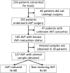
Setting & participants: 127 patients with CKD undergoing AVF surgery at a large academic medical center.
Predictors: Preexisting arterial microcalcification (≥1% of media area) assessed independently by von Kossa stains of arterial specimens obtained during AVF surgery and by preoperative ultrasound.
Outcomes: Juxta-anastomotic AVF stenosis (ascertained by ultrasound obtained 4-6 weeks postoperatively), AVF nonmaturation (inability to cannulate with 2 needles with dialysis blood flow ≥ 300mL/min for ≥6 sessions in 1 month within 6 months of AVF creation), and duration of primary unassisted AVF survival after successful use (time to first intervention).
Results: Arterial microcalcification was present by histologic evaluation in 40% of patients undergoing AVF surgery. The frequency of a postoperative juxta-anastomotic AVF stenosis was similar in patients with or without preexisting arterial microcalcification (32% vs 42%; OR, 0.65; 95% CI, 0.28-1.52; P=0.3). AVF nonmaturation was observed in 29%, 33%, 33%, and 33% of patients with <1%, 1% to 4.9%, 5% to 9.9%, and ≥10% arterial microcalcification, respectively (P=0.9). Sonographic arterial microcalcification was found in 39% of patients and was associated with histologic calcification (P=0.001), but did not predict AVF nonmaturation. Finally, among AVFs that matured, unassisted AVF maturation (time to first intervention) was similar for patients with and without preexisting arterial microcalcification (HR, 0.64; 95% CI, 0.35-1.21; P=0.2).
Limitations: Single-center study.
Conclusions: Arterial microcalcification is common in patients with advanced CKD, but does not explain postoperative AVF stenosis, AVF nonmaturation, or AVF failure after successful cannulation.
Keywords: AVF non-maturation; AVF survival; Arteriovenous fistula (AVF); arterial micro-calcification; arteriovenous access; cannulation; chronic kidney disease (CKD); hemodialysis; juxta-anastomotic AVF stenosis; vascular access; vascular calcification; vascular ultrasound; von Kossa staining.
Copyright © 2015 National Kidney Foundation, Inc. Published by Elsevier Inc. All rights reserved.
Figures


References
-
- National Kidney Foundation: KDOQI clinical practice guidelines and clinical practice recommendations for vascular access 2006. Am J Kidney Dis. 2006;48(suppl 1):S176–S322. - PubMed
-
- Allon M, Robbin ML. Increasing arteriovenous fistulas in hemodialysis patients: problems and solutions. Kidney Int. 2002;62:1109–24. - PubMed
-
- Dember LM, Beck GJ, Allon M, Delmez JA, Dixon BS, Greenberg A, et al. Effect of clopidogrel on early failure of arteriovenous fistulas for hemodialysis. JAMA. 2008;299:2164–71. - PubMed
Publication types
MeSH terms
Grants and funding
LinkOut - more resources
Full Text Sources
Other Literature Sources
Medical

